
Persistent stapedial artery
Citation, DOI, disclosures and article data
At the time the article was created Haris Sair had no recorded disclosures.
View Haris Sair's current disclosuresAt the time the article was last revised Yahya Baba had no financial relationships to ineligible companies to disclose.
View Yahya Baba's current disclosures- Persistent stapedial artery (PSA)
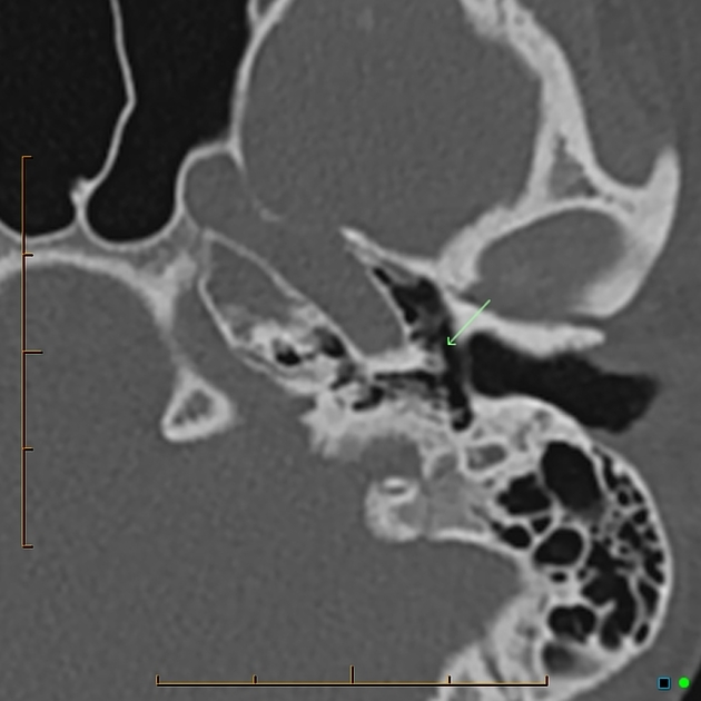
The persistent stapedial artery (PSA) is an abnormal small vessel arising from the petrous portion of the internal carotid artery and crossing through the middle ear. It results from the failure of regression of the embryonic stapedial artery.
On this page:
Epidemiology
The prevalence is thought to range from 0.02 to 0.48% in the population.
Clinical presentation
- pulsatile tinnitus
- conductive hearing loss due to ankylosis of the stapes
- sensorineural hearing loss due to an erosion of otic capsule (rare)
- mass seen over the promontory in the middle ear while performing an otoscopic examination
- incidental finding during surgery that can lead to hemorrhage if not properly identified
Gross anatomy
- arises from the proximal hyoid artery at 4-5 weeks of life
- passes through the obturator foramen of stapes
- branches into upper and lower divisions
- posterior division of the upper branch becomes middle meningeal artery
- lower branch (maxillomandibular) has two branches
- mandibular artery (becomes inferior alveolar artery in adult)
- infraorbital artery (becomes infraorbital artery in adult)
Anastomosis forms between the ventral pharyngeal artery (precursor of external carotid artery) and the lower division of the stapedial artery.
As the ventral pharyngeal artery supplies flow to the middle meningeal artery, stapedial artery regresses, leaving a small caroticotympanic artery.
It is termed persistent as it is normally found until week 10, at which point flow reverses at the foramen spinosum which in turn induces degeneration over the course of the 3rd fetal month.
ADVERTISEMENT: Supporters see fewer/no ads
Blood supply
Can arise from:
Radiographic features
- small canaliculus originating from the petrous segment of the internal carotid artery
- linear soft tissue density crossing over the cochlear promontory
- enlarged facial nerve canal or separate canal parallel to facial nerve
- aplastic or hypoplastic foramen spinosum
- may be a normal variant or in instances where middle meningeal artery arises from the ophthalmic artery
History and etymology
It was first discovered in 1836 at postmortem examination by Austrian anatomist Joseph Hyrtl (1810-1894) 6,7, most famous for his description of the tympanomeningeal fissure.
Related pathology
- may be associated with aberrant internal carotid artery, which is formed when inferior tympanic artery anastomosis with the caroticotympanic artery; enlarged inferior tympanic canaliculus is then seen
- may be associated with other middle ear anomalies
References
- 1. Silbergleit R, Quint DJ, Mehta BA et-al. The persistent stapedial artery. AJNR Am J Neuroradiol. 2000;21 (3): 572-7. AJNR Am J Neuroradiol (full text) - Pubmed citation
- 2. Thiers FA, Sakai O, Poe DS et-al. Persistent stapedial artery: CT findings. AJNR Am J Neuroradiol. 2000;21 (8): 1551-4. AJNR Am J Neuroradiol (full text) - Pubmed citation
- 3. Hatipoglu HG, Cetin MA, Yuksel E et-al. A case of a coexisting aberrant internal carotid artery and persistent stapedial artery: the role of MR angiography in the diagnosis. Ear Nose Throat J. 2011;90 (5): E17-20. Pubmed citation
- 4. Jain R, Gandhi D, Gujar S et-al. Case 67: Persistent stapedial artery. Radiology. 2004;230 (2): 413-6. Radiology (full text) - doi:10.1148/radiol.2302021108 - Pubmed citation
- 5. Yilmaz T, Bilgen C, Savas R et-al. Persistent stapedial artery: MR angiographic and CT findings. AJNR Am J Neuroradiol. 2003;24 (6): 1133-5. AJNR Am J Neuroradiol (full text) - Pubmed citation
- 6. Hyrtl J. Neue beobachtungen aus dem gebiete der menschlichen und vergleichenden anatomie. (1836) Med Jahrb d Oesterreich Staates. 10:457-466
- 7. Tien HC, Linthicum Jr FH. Persistent stapedial artery. (2001) Otology & Neurotology. 22(6):975-6.
Incoming Links
Related articles: Anatomy: Head and neck
- skeleton of the head and neck
-
cranial vault
- scalp (mnemonic)
- fontanelle
-
sutures
- calvarial
- facial
- frontozygomatic suture
- frontomaxillary suture
- frontolacrimal suture
- frontonasal suture
- temporozygomatic suture
- zygomaticomaxillary suture
- parietotemporal suture (parietomastoid suture)
- occipitotemporal suture (occipitomastoid suture)
- sphenofrontal suture
- sphenozygomatic suture
- spheno-occipital suture (not a true suture)
- lacrimomaxillary suture
- nasomaxillary suture
- internasal suture
- basal/internal
- skull landmarks
- frontal bone
- temporal bone
- parietal bone
- occipital bone
- skull base (foramina)
-
facial bones
- midline single bones
- paired bilateral bones
- cervical spine
- hyoid bone
- laryngeal cartilages
-
cranial vault
- muscles of the head and neck
- muscles of the tongue (mnemonic)
- muscles of mastication
-
facial muscles
- epicranius muscle
- circumorbital and palpebral muscles
- nasal muscles
-
buccolabial muscles
- elevators, retractors and evertors of the upper lip
- levator labii superioris alaeque nasalis muscle
- levator labii superioris muscle
- zygomaticus major muscle
- zygomaticus minor muscle
- levator anguli oris muscle
- malaris muscle
- risorius muscle
- depressors, retractors and evertors of the lower lip
- depressor labii inferioris muscle
- depressor anguli oris muscle
- mentalis muscle
- compound sphincter
-
orbicularis oris muscle
- incisivus labii superioris muscle
- incisivus labii inferioris muscle
-
orbicularis oris muscle
- muscle of mastication
- modiolus
- elevators, retractors and evertors of the upper lip
- muscles of the middle ear
- orbital muscles
- muscles of the soft palate
- pharyngeal muscles
- suprahyoid muscles
- infrahyoid muscles
- intrinsic muscles of the larynx
- muscles of the neck
- platysma muscle
- longus colli muscle
- longus capitis muscle
- scalenus anterior muscle
- scalenus medius muscle
- scalenus posterior muscle
- scalenus pleuralis muscle
- sternocleidomastoid muscle
-
suboccipital muscles
- rectus capitis posterior major muscle
- rectus capitis posterior minor muscle
- obliquus capitis superior muscle
- obliquus capitis inferior muscle
- accessory muscles of the neck
- deep cervical fascia
-
deep spaces of the neck
- anterior cervical space
- buccal space
- carotid space
- danger space
- deep cervical fascia
- infratemporal fossa
- masticator space
- parapharyngeal space
- stylomandibular tunnel
- parotid space
- pharyngeal (superficial) mucosal space
- perivertebral space
- posterior cervical space
- pterygopalatine fossa
- retropharyngeal space
- suprasternal space (of Burns)
- visceral space
- surgical triangles of the neck
- orbit
- ear
- paranasal sinuses
- upper respiratory tract
- viscera of the neck
- blood supply of the head and neck
-
arterial supply
-
common carotid artery
- carotid body
- carotid bifurcation
- subclavian artery
- variants
-
common carotid artery
- venous drainage
-
arterial supply
- innervation of the head and neck
-
cranial nerves
- olfactory nerve (CN I)
- optic nerve (CN II)
- oculomotor nerve (CN III)
- trochlear nerve (CN IV)
-
trigeminal nerve (CN V) (mnemonic)
- trigeminal ganglion
- ophthalmic division
- maxillary division
- mandibular division
- abducens nerve (CN VI)
- facial nerve (CN VII)
-
vestibulocochlear nerve (CN VIII)
- vestibular ganglion (Scarpa's ganglion)
- glossopharyngeal nerve (CN IX)
- vagus nerve (CN X)
- (spinal) accessory nerve (CN XI)
- hypoglossal nerve (CN XII)
- parasympathetic ganglia of the head and neck
- cervical sympathetic ganglia
- greater occipital nerve
- third occipital nerve
-
cervical plexus
- muscular branches
- longus capitis
- longus colli
- scalenes
- geniohyoid
- thyrohyoid
-
ansa cervicalis
- omohyoid (superior and inferior bellies separately)
- sternothyroid
- sternohyoid
- phrenic nerve
- contribution to the accessory nerve (CN XI)
- cutaneous branches
- muscular branches
- brachial plexus
- pharyngeal plexus
-
cranial nerves
- lymphatic drainage of the head and neck
- embryological development of the head and neck


 Unable to process the form. Check for errors and try again.