
Pharynx
Citation, DOI, disclosures and article data
At the time the article was created Craig Hacking had no recorded disclosures.
View Craig Hacking's current disclosuresAt the time the article was last revised Craig Hacking had the following disclosures:
- Philips Australia, Paid speaker at Philips Spectral CT events (ongoing)
These were assessed during peer review and were determined to not be relevant to the changes that were made.
View Craig Hacking's current disclosures- Pharynges
- Pharynxes
The pharynx (plural: pharynges or pharynxes) is the superior dilated part of the alimentary tract that connects the nasal and oral cavities to the esophagus. It also forms part of the upper respiratory tract.
On this page:
Gross anatomy
The pharynx is composed of three parts:
-
nasopharynx: posterior to the nasal choanae, extending from the vault of the pharynx superiorly to the soft palate inferiorly
communicates with the nasal cavity anteriorly
-
oropharynx: posterior to the base of tongue, inferior to the soft palate, bounded laterally by the palatoglossal and palatopharyngeal arches, and superior to the superior tip of the epiglottis
communicates with the oral cavity anteriorly
-
laryngopharynx (or hypopharynx): inferior to the superior border of the epiglottis and the pharyngoepiglottic folds, superior to the cricoid cartilage
communicates with the larynx anteriorly
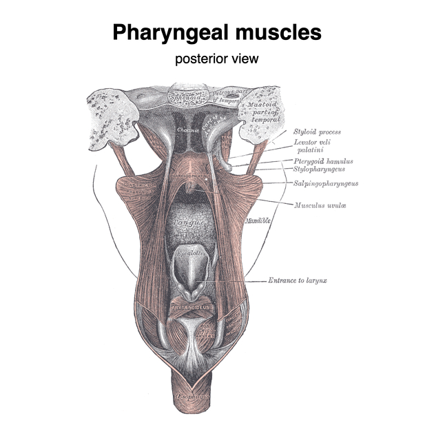
Musculature
There are two groups of pharyngeal muscles, the external circular layer and the internal longitudinal layer.
The external circular layer is composed of the three pharyngeal constrictor muscles:
The internal longitudinal layer is composed of the three paired muscles:
ADVERTISEMENT: Supporters see fewer/no ads
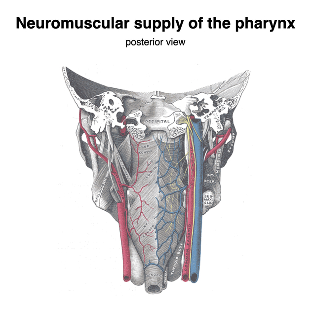
Arterial supply
Numerous branches anastomose in the pharynx, providing it with a rich arterial supply:
ascending pharyngeal artery (from the external carotid artery (ECA))
ascending palatine artery (from the facial artery)
lingual artery (from the ECA)
tonsillar artery (from the facial artery)
greater palatine artery (from the maxillary artery)
artery of the pterygoid canal (from the maxillary artery)
superior laryngeal artery (from the superior thyroid artery)
inferior laryngeal artery (from the inferior thyroid artery, off the thyrocervical trunk)
Venous drainage
Veins of the same name drain either into the pterygoid venous plexus or directly into the internal jugular vein.
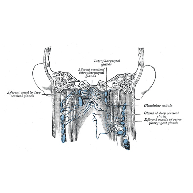
Lymphatic drainage
Most lymph drains back to the retropharyngeal nodes.
Innervation
The muscles of the pharynx are supplied by the pharyngeal plexus, a network of nerves from pharyngeal branches of the vagus and glossopharyngeal nerves.
Sensory innervation is primarily from the glossopharyngeal nerve with a few notable exceptions:
the superior nasopharynx is supplied by the pharyngeal nerve of the maxillary division of the trigeminal nerve (V2)
the valleculae are supplied by the internal laryngeal nerve (a branch of the vagus nerve)
the rest of the laryngopharynx is supplied by the recurrent laryngeal nerve (a branch of the vagus nerve)
References
- 1. McMinn. Last's Anatomy. (2003) ISBN: 9780729537520 - Google Books
- 2. Keith L. Moore, Arthur F. Dalley. Clinically Oriented Anatomy. (1999) ISBN: 9780683061413 - Google Books
Incoming Links
- Vertebral levels (anatomical landmarks)
- Aerodigestive tract
- Spinal nucleus of the trigeminal nerve
- Superior pharyngeal constrictor muscle
- Cervical lung hernia
- Inferior pharyngeal constrictor muscle
- Point-of-care ultrasound (curriculum)
- Nucleus ambiguus
- Oesophageal fibrovascular polyp
- Ariboflavinosis
- Passavant cushion
- Common carotid artery
- Tensor veli palatini muscle
- Pharyngeal muscles
- Nasopharynx
- Oropharynx
- Jugulodigastric lymph nodes
- Esophagus
- Head and neck anatomy
- Respiratory tract
Related articles: Anatomy: Head and neck
- skeleton of the head and neck
-
cranial vault
- scalp (mnemonic)
- fontanelle
-
sutures
- calvarial
- facial
- frontozygomatic suture
- frontomaxillary suture
- frontolacrimal suture
- frontonasal suture
- temporozygomatic suture
- zygomaticomaxillary suture
- parietotemporal suture (parietomastoid suture)
- occipitotemporal suture (occipitomastoid suture)
- sphenofrontal suture
- sphenozygomatic suture
- spheno-occipital suture (not a true suture)
- lacrimomaxillary suture
- nasomaxillary suture
- internasal suture
- basal/internal
- skull landmarks
- frontal bone
- temporal bone
- parietal bone
- occipital bone
- skull base (foramina)
-
facial bones
- midline single bones
- paired bilateral bones
- cervical spine
- hyoid bone
- laryngeal cartilages
-
cranial vault
- muscles of the head and neck
- muscles of the tongue (mnemonic)
- muscles of mastication
-
facial muscles
- epicranius muscle
- circumorbital and palpebral muscles
- nasal muscles
-
buccolabial muscles
- elevators, retractors and evertors of the upper lip
- levator labii superioris alaeque nasalis muscle
- levator labii superioris muscle
- zygomaticus major muscle
- zygomaticus minor muscle
- levator anguli oris muscle
- malaris muscle
- risorius muscle
- depressors, retractors and evertors of the lower lip
- depressor labii inferioris muscle
- depressor anguli oris muscle
- mentalis muscle
- compound sphincter
-
orbicularis oris muscle
- incisivus labii superioris muscle
- incisivus labii inferioris muscle
-
orbicularis oris muscle
- muscle of mastication
- modiolus
- elevators, retractors and evertors of the upper lip
- muscles of the middle ear
- orbital muscles
- muscles of the soft palate
- pharyngeal muscles
- suprahyoid muscles
- infrahyoid muscles
- intrinsic muscles of the larynx
- muscles of the neck
- platysma muscle
- longus colli muscle
- longus capitis muscle
- scalenus anterior muscle
- scalenus medius muscle
- scalenus posterior muscle
- scalenus pleuralis muscle
- sternocleidomastoid muscle
-
suboccipital muscles
- rectus capitis posterior major muscle
- rectus capitis posterior minor muscle
- obliquus capitis superior muscle
- obliquus capitis inferior muscle
- accessory muscles of the neck
- deep cervical fascia
-
deep spaces of the neck
- anterior cervical space
- buccal space
- carotid space
- danger space
- deep cervical fascia
- infratemporal fossa
- masticator space
- parapharyngeal space
- stylomandibular tunnel
- parotid space
- pharyngeal (superficial) mucosal space
- perivertebral space
- posterior cervical space
- pterygopalatine fossa
- retropharyngeal space
- suprasternal space (of Burns)
- visceral space
- surgical triangles of the neck
- orbit
- ear
- paranasal sinuses
- upper respiratory tract
- viscera of the neck
- blood supply of the head and neck
-
arterial supply
-
common carotid artery
- carotid body
- carotid bifurcation
- subclavian artery
- variants
-
common carotid artery
- venous drainage
-
arterial supply
- innervation of the head and neck
-
cranial nerves
- olfactory nerve (CN I)
- optic nerve (CN II)
- oculomotor nerve (CN III)
- trochlear nerve (CN IV)
-
trigeminal nerve (CN V) (mnemonic)
- trigeminal ganglion
- ophthalmic division
- maxillary division
- mandibular division
- abducens nerve (CN VI)
- facial nerve (CN VII)
-
vestibulocochlear nerve (CN VIII)
- vestibular ganglion (Scarpa's ganglion)
- glossopharyngeal nerve (CN IX)
- vagus nerve (CN X)
- (spinal) accessory nerve (CN XI)
- hypoglossal nerve (CN XII)
- parasympathetic ganglia of the head and neck
- cervical sympathetic ganglia
- greater occipital nerve
- third occipital nerve
-
cervical plexus
- muscular branches
- longus capitis
- longus colli
- scalenes
- geniohyoid
- thyrohyoid
-
ansa cervicalis
- omohyoid (superior and inferior bellies separately)
- sternothyroid
- sternohyoid
- phrenic nerve
- contribution to the accessory nerve (CN XI)
- cutaneous branches
- muscular branches
- brachial plexus
- pharyngeal plexus
-
cranial nerves
- lymphatic drainage of the head and neck
- embryological development of the head and neck


 Unable to process the form. Check for errors and try again.