
Choroidal fissure
Citation, DOI, disclosures and article data
At the time the article was created Maxime St-Amant had no recorded disclosures.
View Maxime St-Amant's current disclosuresAt the time the article was last revised Yvette Mellam had no financial relationships to ineligible companies to disclose.
View Yvette Mellam's current disclosures- Choroid fissure
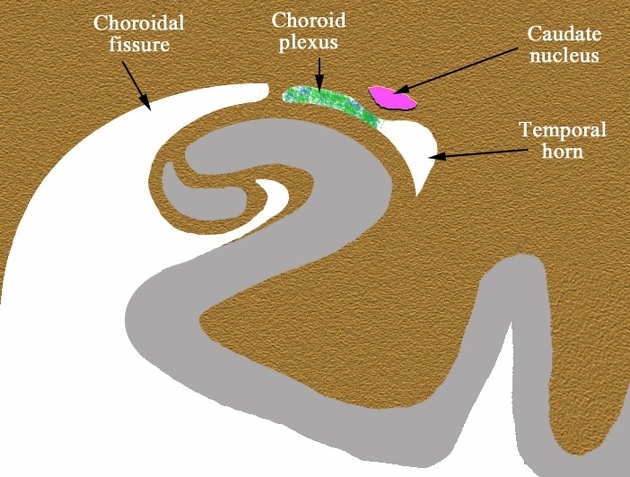
The choroidal fissure, or choroid fissure, is a cleft of the medial surface of the cerebral hemisphere running immediately above the hippocampus and extends around the thalamus to the interventricular foramen of Monroe. It forms the medial wall of the lateral ventricle and attachment site for the choroid plexus.
Gross anatomy
The choroidal fissue is a C-shaped fissure in the medial wall of the cerebral hemisphere; extending from the interventricular foramen of Monroe, to around the thalamus and cerebral peduncle as far as the uncus of the temporal lobe.
The convexity of the C shape is contained by the body and crus (pillar) of the fornix, the fimbria and the hippocampus.
The concavity of the C shaped slit is contained by the thalamus (upper and posterior surfaces) and the tail of the caudate nucleus.
The slit of the choroid fissure is where pia mater and ependyma are in direct contact with each other; evaginating into the lateral ventricles as choroid plexus1.
Opening the choroidal fissue between the fornix and the thalamus will lead into the velum interpositum.
At the level of the hippocampus, the choroidal fissure is a lateral extension of the transverse fissure of Bichat, which in turn is a lateral extension of the ambient cistern 4,5.
The choroid plexus originates at the inferior choroidal point of the choroidal fissure, which is located just posterior to the hippocampal head. At this location, the anterior choroidal artery enters the temporal horn of the lateral ventricle. Only choroid plexus and a thin arachnoid membrane separate the cerebrospinal fluid space of the choroidal fissure and the lateral ventricle.
ADVERTISEMENT: Supporters see fewer/no ads
Related pathology

References
- 1. Robert M. H. McMinn. Last's Anatomy. (2019) ISBN: 9780729543576 - Google Books
- 4. Holodny AI, George AE, Golomb J, de Leon MJ, Kalnin AJ. The perihippocampal fissures: normal anatomy and disease states. (1998) Radiographics. 18 (3): 653-65. doi:10.1148/radiographics.18.3.9599389 - Pubmed
- 5. Dekeyzer S, De Kock I, Nikoubashman O, Vanden Bossche S, Van Eetvelde R, De Groote J, Acou M, Wiesmann M, Deblaere K, Achten E. "Unforgettable" - a pictorial essay on anatomy and pathology of the hippocampus. (2017) Insights into imaging. 8 (2): 199-212. doi:10.1007/s13244-016-0541-2 - Pubmed
Incoming Links
Related articles: Anatomy: Brain
-
brain
- grey matter
- white matter
-
cerebrum
-
cerebral hemisphere (telencephalon)
- cerebral lobes and gyri
- frontal lobe
- parietal lobe
-
occipital lobe
- occipital pole
- lingual gyrus
- fusiform gyrus (Brodmann area 37)
- calcarine (visual) cortex
- cuneus
- temporal lobe
- basal forebrain
- limbic system
- insula
-
cerebral sulci and fissures (A-Z)
- calcarine fissure
- callosal sulcus
- central (Rolandic) sulcus
- cingulate sulcus
- collateral sulcus
- inferior frontal sulcus
- inferior occipital sulcus
- inferior temporal sulcus
- interhemispheric fissure
- intraparietal sulcus
- lateral (Sylvian) sulcus
- lateral occipital sulcus
- marginal sulcus
- occipitotemporal sulcus
- olfactory sulcus
- paracentral sulcus
- paraolfactory sulcus
- parieto-occipital fissure
- posterior parolfactory sulcus
- precentral sulcus
- preoccipital notch
- postcentral sulcus
- rhinal sulcus
- rostral sulcus
- subparietal sulcus
- superior frontal sulcus
- superior occipital sulcus
- superior temporal sulcus
- cortical histology
- cerebral lobes and gyri
- white matter tracts
- deep grey matter
-
pituitary gland
- posterior pituitary and stalk (part of diencephalon)
- anterior pituitary
- inferior hypophyseal arterial circle
- diencephalon
-
cerebral hemisphere (telencephalon)
-
brainstem
- midbrain (mesencephalon)
- pons (part of metencephalon)
- medulla oblongata (myelencephalon)
- white matter
-
grey matter
- non-cranial nerve
-
cranial nerve nuclei
- oculomotor nucleus
- Edinger-Westphal nucleus
- trochlear nucleus
- motor nucleus of CN V
- mesencephalic nucleus of CN V
- main sensory nucleus of CN V
- spinal nucleus of CN V
- abducent nucleus
- facial nucleus
- superior salivatory nucleus
- cochlear nuclei
- vestibular nuclei
- inferior salivatory nucleus
- solitary tract nucleus
- ambiguus nucleus
- dorsal vagal motor nucleus
- hypoglossal nucleus
-
cerebellum (part of metencephalon)
- vermis
- cerebellar hemisphere
- cerebellar peduncles
- cranial meninges (meninx primitiva)
- CSF spaces
-
cranial nerves (mnemonic)
- olfactory nerve (CN I)
- optic nerve (CN II)
- oculomotor nerve (CN III)
- trochlear nerve (CN IV)
- trigeminal nerve (CN V) (mnemonic)
- abducens nerve (CN VI)
- facial nerve (CN VII) (segments mnemonic | branches mnemonic)
-
vestibulocochlear nerve (CN VIII)
- vestibular ganglion (Scarpa's ganglion)
- glossopharyngeal nerve (CN IX)
- vagus nerve (CN X)
- spinal accessory nerve (CN XI)
- hypoglossal nerve (CN XII)
- functional neuroanatomy
- CNS development
- cerebral vascular supply
- arteries
- vascular territories
-
circle of Willis
- internal carotid artery (ICA) (segments)
- vertebral artery
-
normal variants
- intracranial arterial fenestration
- internal carotid artery (ICA)
- anterior cerebral artery (ACA)
- middle cerebral artery (MCA)
- posterior cerebral artery (PCA)
- basilar artery
- persistent carotid-vertebrobasilar artery anastomoses (mnemonic)
- vertebral artery
- ophthalmic artery
-
cerebral venous system
-
dural venous sinuses
- basilar venous plexus
- cavernous sinus (mnemonic)
- clival diploic veins
- inferior petro-occipital vein
- inferior petrosal sinus
- inferior sagittal sinus
- intercavernous sinus
- internal carotid artery venous plexus of Rektorzik
- jugular bulb
- marginal sinus
- occipital sinus
- sigmoid sinus
- sphenoparietal sinus
- straight sinus
- superior petrosal sinus
- superior sagittal sinus
- torcula herophili
- transverse sinus
-
cerebral veins
-
superficial veins of the brain
- superior cerebral veins (superficial cerebral veins)
- inferior cerebral veins
- superficial middle cerebral vein
- superior anastomotic vein (of Trolard)
- inferior anastomotic vein (of Labbe)
-
superficial veins of the brain
-
deep veins of the brain
- great cerebral vein (of Galen)
- venous circle of Trolard
- normal variants
-
dural venous sinuses
- arteries
- glymphatic pathway


 Unable to process the form. Check for errors and try again.