
Main sensory nucleus of the trigeminal nerve
Citation, DOI, disclosures and article data
At the time the article was created Chris Rothe had no recorded disclosures.
View Chris Rothe's current disclosuresAt the time the article was last revised Rohit Sharma had no financial relationships to ineligible companies to disclose.
View Rohit Sharma's current disclosures- main sensory nucleus
- chief sensory nucleus
- chief sensory nucleus of the trigeminal nerve
- main trigeminal sensory nucleus
- chief trigeminal sensory nucleus
- pontine trigeminal nucleus
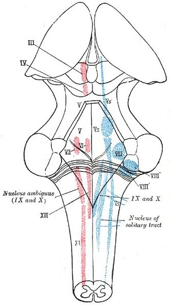
The main or chief sensory nucleus of the trigeminal nerve is one of three major nuclei that make up the trigeminal sensory nerve nuclear complex along with the mesencephalic nucleus and the spinal nucleus 1,2. It also receives fibers from the glossopharyngeal nerve.
Some older texts refer to it as the pontine trigeminal nucleus due to its location.
Gross anatomy
The main sensory nucleus of the trigeminal nerve is located within the caudal aspect of the pons. It lies inferior to the mesencephalic nucleus and superior to the spinal nucleus and is continuous with both. It lies dorsomedial to the entering trigeminal root and also posterolateral to the motor trigeminal nucleus.
Innervation
The main sensory nucleus receives facial tactile sensation in addition to receiving proprioceptive input from the temporomandibular joint.
The nucleus of the trigeminal nerve is conventionally divided into dorsomedial and ventrolateral divisions 2. The dorsomedial division receives afferent input from the oral cavity. The ventrolateral division receives input from all three divisions (V1-V3) of the trigeminal nerve.
References
- 1. Lynley Bradnam, Christine Barry. The Role of the Trigeminal Sensory Nuclear Complex in the Pathophysiology of Craniocervical Dystonia. (2013) Journal of Neuroscience. 33 (47): 18358. doi:10.1523/JNEUROSCI.3544-13.2013 - Pubmed
- 2. Shmuel Price, Daniel T. Daly. Neuroanatomy, Nucleus Trigeminal. (2019) Pubmed
Incoming Links
Related articles: Anatomy: Brain
-
brain
- grey matter
- white matter
-
cerebrum [+][+]
-
cerebral hemisphere (telencephalon)
- cerebral lobes and gyri
- frontal lobe
- parietal lobe
-
occipital lobe
- occipital pole
- lingual gyrus
- fusiform gyrus (Brodmann area 37)
- calcarine (visual) cortex
- cuneus
- temporal lobe
- basal forebrain
- limbic system
- insula
-
cerebral sulci and fissures (A-Z)
- calcarine fissure
- callosal sulcus
- central (Rolandic) sulcus
- cingulate sulcus
- collateral sulcus
- inferior frontal sulcus
- inferior occipital sulcus
- inferior temporal sulcus
- interhemispheric fissure
- intraparietal sulcus
- lateral (Sylvian) sulcus
- lateral occipital sulcus
- marginal sulcus
- occipitotemporal sulcus
- olfactory sulcus
- paracentral sulcus
- paraolfactory sulcus
- parieto-occipital fissure
- posterior parolfactory sulcus
- precentral sulcus
- preoccipital notch
- postcentral sulcus
- rhinal sulcus
- rostral sulcus
- subparietal sulcus
- superior frontal sulcus
- superior occipital sulcus
- superior temporal sulcus
- cortical histology
- cerebral lobes and gyri
- white matter tracts
- deep grey matter
-
pituitary gland
- posterior pituitary and stalk (part of diencephalon)
- anterior pituitary
- inferior hypophyseal arterial circle
- diencephalon
-
cerebral hemisphere (telencephalon)
-
brainstem
- midbrain (mesencephalon)[+][+]
- pons (part of metencephalon)[+][+]
- medulla oblongata (myelencephalon)[+][+]
- white matter
-
grey matter
- non-cranial nerve[+][+]
-
cranial nerve nuclei
- oculomotor nucleus
- Edinger-Westphal nucleus
- trochlear nucleus
- motor nucleus of CN V
- mesencephalic nucleus of CN V
- main sensory nucleus of CN V
- spinal nucleus of CN V
- abducent nucleus
- facial nucleus
- superior salivatory nucleus
- cochlear nuclei
- vestibular nuclei
- inferior salivatory nucleus
- solitary tract nucleus
- ambiguus nucleus
- dorsal vagal motor nucleus
- hypoglossal nucleus
-
cerebellum (part of metencephalon)[+][+]
- vermis
- cerebellar hemisphere
- cerebellar peduncles
- cranial meninges (meninx primitiva)[+][+]
- CSF spaces[+][+]
-
cranial nerves (mnemonic)[+][+]
- olfactory nerve (CN I)
- optic nerve (CN II)
- oculomotor nerve (CN III)
- trochlear nerve (CN IV)
- trigeminal nerve (CN V) (mnemonic)
- abducens nerve (CN VI)
- facial nerve (CN VII) (segments mnemonic | branches mnemonic)
-
vestibulocochlear nerve (CN VIII)
- vestibular ganglion (Scarpa's ganglion)
- glossopharyngeal nerve (CN IX)
- vagus nerve (CN X)
- spinal accessory nerve (CN XI)
- hypoglossal nerve (CN XII)
- functional neuroanatomy[+][+]
- CNS development[+][+]
- cerebral vascular supply[+][+]
- arteries
- vascular territories
-
circle of Willis
- internal carotid artery (ICA) (segments)
- vertebral artery
-
normal variants
- intracranial arterial fenestration
- internal carotid artery (ICA)
- anterior cerebral artery (ACA)
- middle cerebral artery (MCA)
- posterior cerebral artery (PCA)
- basilar artery
- persistent carotid-vertebrobasilar artery anastomoses (mnemonic)
- vertebral artery
- ophthalmic artery
-
cerebral venous system
-
dural venous sinuses
- basilar venous plexus
- cavernous sinus (mnemonic)
- clival diploic veins
- inferior petro-occipital vein
- inferior petrosal sinus
- inferior sagittal sinus
- intercavernous sinus
- internal carotid artery venous plexus of Rektorzik
- jugular bulb
- marginal sinus
- occipital sinus
- sigmoid sinus
- sphenoparietal sinus
- straight sinus
- superior petrosal sinus
- superior sagittal sinus
- torcula herophili
- transverse sinus
-
cerebral veins
-
superficial veins of the brain
- superior cerebral veins (superficial cerebral veins)
- inferior cerebral veins
- superficial middle cerebral vein
- superior anastomotic vein (of Trolard)
- inferior anastomotic vein (of Labbe)
-
superficial veins of the brain
-
deep veins of the brain
- great cerebral vein (of Galen)
- venous circle of Trolard
- normal variants
-
dural venous sinuses
- arteries
- glymphatic pathway


 Unable to process the form. Check for errors and try again.