
Hypodontia
Citation, DOI, disclosures and article data
At the time the article was created Henry Knipe had no recorded disclosures.
View Henry Knipe's current disclosuresAt the time the article was last revised Daniel J Bell had no financial relationships to ineligible companies to disclose.
View Daniel J Bell's current disclosures- Oligodontia
- Adontia
- Congenitally missing teeth
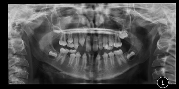
Hypodontia, also known as oligodontia or adontia, refers to the developmental failure of one or more teeth, excluding the third molars.
On this page:
Terminology
The phenomenon of non-development of teeth has been described using several terms including 'hypodontia', 'oligodontia' and 'adontia'. The descriptor 'congenitally missing teeth' has also been used, but may be misleading as teeth development is completed following birth so identification is only possible in early childhood.
Epidemiology
Hypodontia is common, affecting between 1.6 to 6.9% of the population with a recognized variations in ethnicities. There is a female preponderance with a M:F of 2:3 1,2.
Pathology
Hypodontia is relatively rare in the primary dentition, mostly affecting the secondary dentition. The maxillary lateral incisors or mandibular premolars are most commonly missing, and in most individuals only one or two teeth are absent . Hypodontia is also associated with microdontia and delayed dental development 1,2.
Absence of one or more of the third molars affects around 20% of the population and is considered part of normal variation rather than hypodontia.
Hypodontia is known to occur with a large range of syndromes including cleft lip and palate, Down syndrome and ectodermal dysplasia, but is also common in non-syndromic patients 1.
Radiographic features
Hypodontia is usually assessed using orthopantomography, although cone beam CT may be utilized for problem solving.
Care should be taken not to confuse late calcification of the developing tooth bud with its absence and some authors suggest avoiding the diagnosis of hypodontia in children younger than 9 or 10 years old 3.
References
- 1. Al-Ani A, Antoun J, Thomson W, Merriman T, Farella M. Hypodontia: An Update on Its Etiology, Classification, and Clinical Management. BioMed Research International. 2017;2017:1-9. doi:10.1155/2017/9378325 - Pubmed
- 2. Valle AL, Lorenzoni FC, Martins LM et-al. A multidisciplinary approach for the management of hypodontia: case report. J Appl Oral Sci. 2012;19 (5): 544-8. Free text at pubmed - Pubmed citation
- 3. Rakhshan V. Congenitally Missing Teeth (Hypodontia): A Review of the Literature Concerning the Etiology, Prevalence, Risk Factors, Patterns and Treatment. Dent Res J. 2015;12(1):1. doi:10.4103/1735-3327.150286 - Pubmed
Incoming Links
Related articles: Anatomy: Head and neck
- skeleton of the head and neck
-
cranial vault
- scalp (mnemonic)
- fontanelle
-
sutures
- calvarial
- facial
- frontozygomatic suture
- frontomaxillary suture
- frontolacrimal suture
- frontonasal suture
- temporozygomatic suture
- zygomaticomaxillary suture
- parietotemporal suture (parietomastoid suture)
- occipitotemporal suture (occipitomastoid suture)
- sphenofrontal suture
- sphenozygomatic suture
- spheno-occipital suture (not a true suture)
- lacrimomaxillary suture
- nasomaxillary suture
- internasal suture
- basal/internal
- skull landmarks
- frontal bone
- temporal bone
- parietal bone
- occipital bone
- skull base (foramina)
-
facial bones
- midline single bones
- paired bilateral bones
- cervical spine
- hyoid bone
- laryngeal cartilages
-
cranial vault
- muscles of the head and neck
- muscles of the tongue (mnemonic)
- muscles of mastication
-
facial muscles
- epicranius muscle
- circumorbital and palpebral muscles
- nasal muscles
-
buccolabial muscles
- elevators, retractors and evertors of the upper lip
- levator labii superioris alaeque nasalis muscle
- levator labii superioris muscle
- zygomaticus major muscle
- zygomaticus minor muscle
- levator anguli oris muscle
- malaris muscle
- risorius muscle
- depressors, retractors and evertors of the lower lip
- depressor labii inferioris muscle
- depressor anguli oris muscle
- mentalis muscle
- compound sphincter
-
orbicularis oris muscle
- incisivus labii superioris muscle
- incisivus labii inferioris muscle
-
orbicularis oris muscle
- muscle of mastication
- modiolus
- elevators, retractors and evertors of the upper lip
- muscles of the middle ear
- orbital muscles
- muscles of the soft palate
- pharyngeal muscles
- suprahyoid muscles
- infrahyoid muscles
- intrinsic muscles of the larynx
- muscles of the neck
- platysma muscle
- longus colli muscle
- longus capitis muscle
- scalenus anterior muscle
- scalenus medius muscle
- scalenus posterior muscle
- scalenus pleuralis muscle
- sternocleidomastoid muscle
-
suboccipital muscles
- rectus capitis posterior major muscle
- rectus capitis posterior minor muscle
- obliquus capitis superior muscle
- obliquus capitis inferior muscle
- accessory muscles of the neck
- deep cervical fascia
-
deep spaces of the neck
- anterior cervical space
- buccal space
- carotid space
- danger space
- deep cervical fascia
- infratemporal fossa
- masticator space
- parapharyngeal space
- stylomandibular tunnel
- parotid space
- pharyngeal (superficial) mucosal space
- perivertebral space
- posterior cervical space
- pterygopalatine fossa
- retropharyngeal space
- suprasternal space (of Burns)
- visceral space
- surgical triangles of the neck
- orbit
- ear
- paranasal sinuses
- upper respiratory tract
- viscera of the neck
- blood supply of the head and neck
-
arterial supply
-
common carotid artery
- carotid body
- carotid bifurcation
- subclavian artery
- variants
-
common carotid artery
- venous drainage
-
arterial supply
- innervation of the head and neck
-
cranial nerves
- olfactory nerve (CN I)
- optic nerve (CN II)
- oculomotor nerve (CN III)
- trochlear nerve (CN IV)
-
trigeminal nerve (CN V) (mnemonic)
- trigeminal ganglion
- ophthalmic division
- maxillary division
- mandibular division
- abducens nerve (CN VI)
- facial nerve (CN VII)
-
vestibulocochlear nerve (CN VIII)
- vestibular ganglion (Scarpa's ganglion)
- glossopharyngeal nerve (CN IX)
- vagus nerve (CN X)
- (spinal) accessory nerve (CN XI)
- hypoglossal nerve (CN XII)
- parasympathetic ganglia of the head and neck
- cervical sympathetic ganglia
- greater occipital nerve
- third occipital nerve
-
cervical plexus
- muscular branches
- longus capitis
- longus colli
- scalenes
- geniohyoid
- thyrohyoid
-
ansa cervicalis
- omohyoid (superior and inferior bellies separately)
- sternothyroid
- sternohyoid
- phrenic nerve
- contribution to the accessory nerve (CN XI)
- cutaneous branches
- muscular branches
- brachial plexus
- pharyngeal plexus
-
cranial nerves
- lymphatic drainage of the head and neck
- embryological development of the head and neck


 Unable to process the form. Check for errors and try again.