
Parietal bone
Citation, DOI, disclosures and article data
At the time the article was created Aaron Wong had no recorded disclosures.
View Aaron Wong's current disclosuresAt the time the article was last revised Joachim Feger had no financial relationships to ineligible companies to disclose.
View Joachim Feger's current disclosures- Os parietale
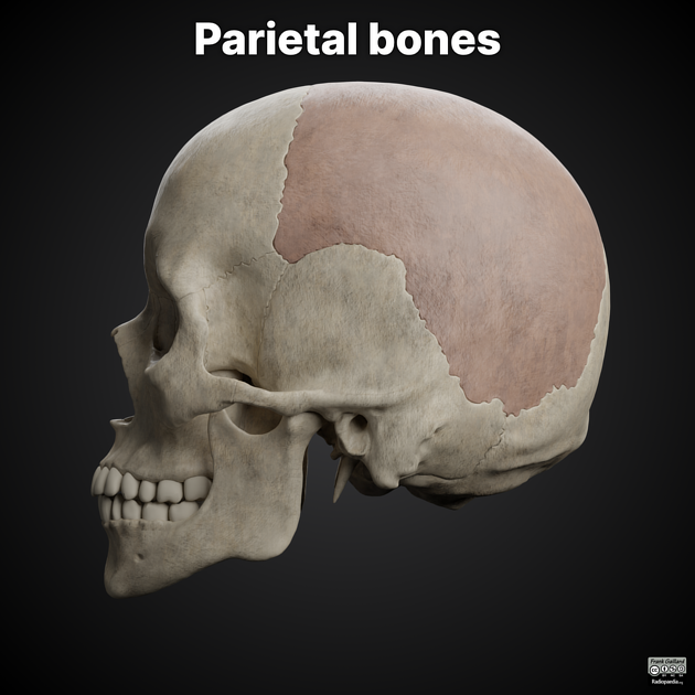
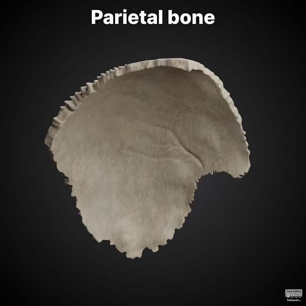
The parietal bone is a paired, irregular, quadrilateral skull bone that forms the sides and roof of the cranium.
On this page:
Gross anatomy
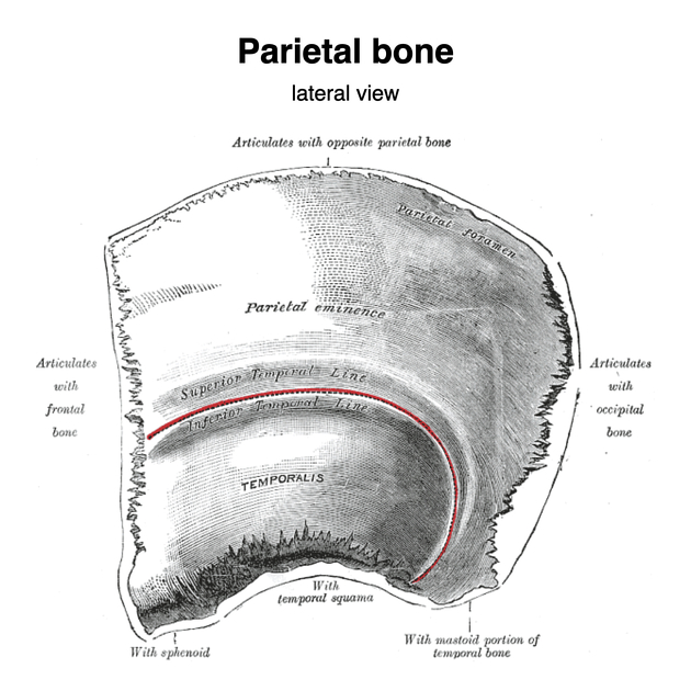
The parietal bone has four borders, four angles, and external/internal surfaces.
The four borders are:
frontal
sagittal
occipital (half of lambdoid suture)
squamous temporal
The four angles are:
sphenoid
mastoid
occipital
frontal
The external surface is convex and smooth and has several features:
parietal eminence
superior temporal line for attachment of the temporalis fascia
inferior temporal lines for attachment of temporalis muscle
parietal foramen contains an emissary vein to the superior sagittal sinus
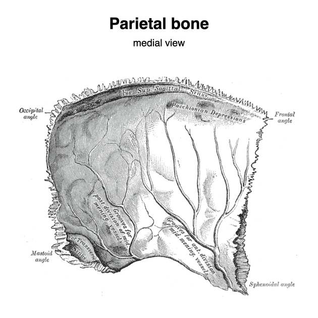
The internal surface is concave and contains grooves for the middle meningeal vessels and the sagittal sulcus.
Articulations
The parietal bone articulates with five other bones:
the frontal bone via the coronal suture
the temporal bone via the squamous suture
the occipital bone via the lambdoid suture
the contralateral parietal bone via the sagittal suture
Anatomical variants
Related pathology
References
- 1. Henry Gray, Patricia Collins. Gray's Anatomy. (2005) ISBN: 0443071683 - Google Books
Incoming Links
- Parietotemporal suture
- Coronal suture
- Middle meningeal artery
- Tentorium cerebelli
- Parietal foramen
- Intramembranous ossification
- Mastoid part of temporal bone
- Greater wing of sphenoid
- Squamous part of temporal bone
- Transcranial Doppler sonography (ultrasound)
- Lambdoid suture
- Accessory sutures
- Base of the skull
- Longitudinal temporal bone fractures
- Skull vault osteoma
- Skull vault bones (mnemonic)
- Axial skeleton
- Parietal lobe
- Squamosal suture
- Occipital bone
Related articles: Anatomy: Head and neck
- skeleton of the head and neck
-
cranial vault
- scalp (mnemonic)
- fontanelle
-
sutures
- calvarial
- facial
- frontozygomatic suture
- frontomaxillary suture
- frontolacrimal suture
- frontonasal suture
- temporozygomatic suture
- zygomaticomaxillary suture
- parietotemporal suture (parietomastoid suture)
- occipitotemporal suture (occipitomastoid suture)
- sphenofrontal suture
- sphenozygomatic suture
- spheno-occipital suture (not a true suture)
- lacrimomaxillary suture
- nasomaxillary suture
- internasal suture
- basal/internal
- skull landmarks
- frontal bone
- temporal bone
- parietal bone
- occipital bone
- skull base (foramina)
-
facial bones
- midline single bones
- paired bilateral bones
- cervical spine
- hyoid bone
- laryngeal cartilages
-
cranial vault
- muscles of the head and neck
- muscles of the tongue (mnemonic)
- muscles of mastication
-
facial muscles
- epicranius muscle
- circumorbital and palpebral muscles
- nasal muscles
-
buccolabial muscles
- elevators, retractors and evertors of the upper lip
- levator labii superioris alaeque nasalis muscle
- levator labii superioris muscle
- zygomaticus major muscle
- zygomaticus minor muscle
- levator anguli oris muscle
- malaris muscle
- risorius muscle
- depressors, retractors and evertors of the lower lip
- depressor labii inferioris muscle
- depressor anguli oris muscle
- mentalis muscle
- compound sphincter
-
orbicularis oris muscle
- incisivus labii superioris muscle
- incisivus labii inferioris muscle
-
orbicularis oris muscle
- muscle of mastication
- modiolus
- elevators, retractors and evertors of the upper lip
- muscles of the middle ear
- orbital muscles
- muscles of the soft palate
- pharyngeal muscles
- suprahyoid muscles
- infrahyoid muscles
- intrinsic muscles of the larynx
- muscles of the neck
- platysma muscle
- longus colli muscle
- longus capitis muscle
- scalenus anterior muscle
- scalenus medius muscle
- scalenus posterior muscle
- scalenus pleuralis muscle
- sternocleidomastoid muscle
-
suboccipital muscles
- rectus capitis posterior major muscle
- rectus capitis posterior minor muscle
- obliquus capitis superior muscle
- obliquus capitis inferior muscle
- accessory muscles of the neck
- deep cervical fascia
-
deep spaces of the neck
- anterior cervical space
- buccal space
- carotid space
- danger space
- deep cervical fascia
- infratemporal fossa
- masticator space
- parapharyngeal space
- stylomandibular tunnel
- parotid space
- pharyngeal (superficial) mucosal space
- perivertebral space
- posterior cervical space
- pterygopalatine fossa
- retropharyngeal space
- suprasternal space (of Burns)
- visceral space
- surgical triangles of the neck
- orbit
- ear
- paranasal sinuses
- upper respiratory tract
- viscera of the neck
- blood supply of the head and neck
-
arterial supply
-
common carotid artery
- carotid body
- carotid bifurcation
- subclavian artery
- variants
-
common carotid artery
- venous drainage
-
arterial supply
- innervation of the head and neck
-
cranial nerves
- olfactory nerve (CN I)
- optic nerve (CN II)
- oculomotor nerve (CN III)
- trochlear nerve (CN IV)
-
trigeminal nerve (CN V) (mnemonic)
- trigeminal ganglion
- ophthalmic division
- maxillary division
- mandibular division
- abducens nerve (CN VI)
- facial nerve (CN VII)
-
vestibulocochlear nerve (CN VIII)
- vestibular ganglion (Scarpa's ganglion)
- glossopharyngeal nerve (CN IX)
- vagus nerve (CN X)
- (spinal) accessory nerve (CN XI)
- hypoglossal nerve (CN XII)
- parasympathetic ganglia of the head and neck
- cervical sympathetic ganglia
- greater occipital nerve
- third occipital nerve
-
cervical plexus
- muscular branches
- longus capitis
- longus colli
- scalenes
- geniohyoid
- thyrohyoid
-
ansa cervicalis
- omohyoid (superior and inferior bellies separately)
- sternothyroid
- sternohyoid
- phrenic nerve
- contribution to the accessory nerve (CN XI)
- cutaneous branches
- muscular branches
- brachial plexus
- pharyngeal plexus
-
cranial nerves
- lymphatic drainage of the head and neck
- embryological development of the head and neck


 Unable to process the form. Check for errors and try again.