
The petrous part of the temporal bone, also known as the petrous temporal bone (PTB), petrous pyramid, or petrous face 5, forms the part of skull base between the sphenoid and occipital bones.
On this page:
Gross anatomy
The petrous temporal bone has a pyramidal shape with an apex and a base as well as three surfaces and angles:
-
apex (petrous apex)
directed medially; articulates with the posterior aspect of the greater wing of the sphenoid and basilar occiput
forms internal border of the carotid canal and the posterolateral boundary of the foramen lacerum
-
base
directed laterally and fuses with the internal surface of squama temporalis and mastoid
The petrous temporal bone has three surfaces - anterior, posterior and inferior.
The anterior surface forms the posterior part of the middle cranial fossa. Laterally, it is continuous with the inner surface of the squamous part united by the petrosquamous suture. Near its center lies the arcuate eminence, which indicates the location of the superior semicircular canal. Lateral to the arcuate eminence is tegmen tympani, where there is a depression at the position of middle ear cavity. A shallow groove directed posterolaterally to open into the hiatus of the facial canal. Lateral to this hiatus a smaller hiatus for the lesser petrosal nerve. Medially, there is the trigeminal impression, which is the location of Meckel's cave. At the apex, the termination of the carotid canal is present.
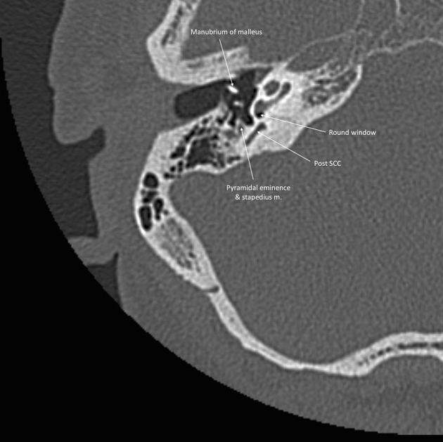
The posterior surface forms the anterior part of posterior cranial fossa. It fuses with the inner surface of the mastoid. Near the center of the posterior surface is the internal acoustic meatus. Posteriorly to the internal acoustic meatus is a small slit, leading to the canal of the vestibular aqueduct.
The inferior surface forms part of the exterior of the base of the skull. There are a number of foramina including the inferior opening of the carotid canal and posteriorly the jugular foramen and in between the petrosal fossula and inferior tympanic canaliculus, through which the tympanic branch of the glossopharyngeal nerve passes. The stylomastoid foramen is situated on the inferior surface. It provides attachment to the levator veli palatini and the cartilaginous portion of the Eustachian tube.
The petrous temporal bone has three angles:
the superior angle with an attachment to the tentorium cerebelli, its medial arm lodges the trigeminal nerve and the superior petrosal sinus lodges in the groove of the angle
the posterior angle which contains a sulcus that houses the inferior petrosal sinus medially, and the jugular notch of the occipital bone forms the jugular foramen laterally
the anterior angle whose medial half articulates with the spinous process of the sphenoid and lateral half fuses with the squamous part at the petrosquamous suture
Variant anatomy
Radiographic features
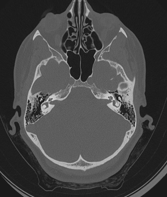
The petrous temporal bone is primarily imaged with CT which is able to provide high resolution bony anatomy sufficient to visualize the inner ear structures and ossicles.
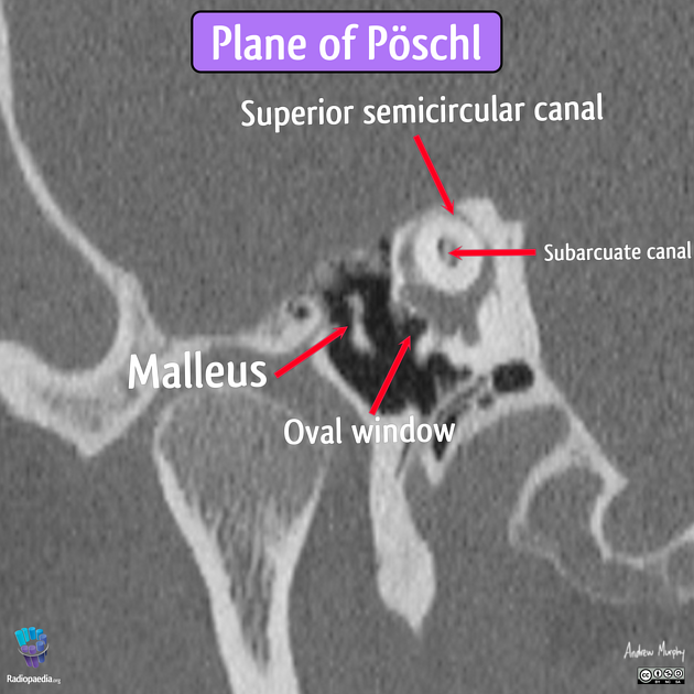
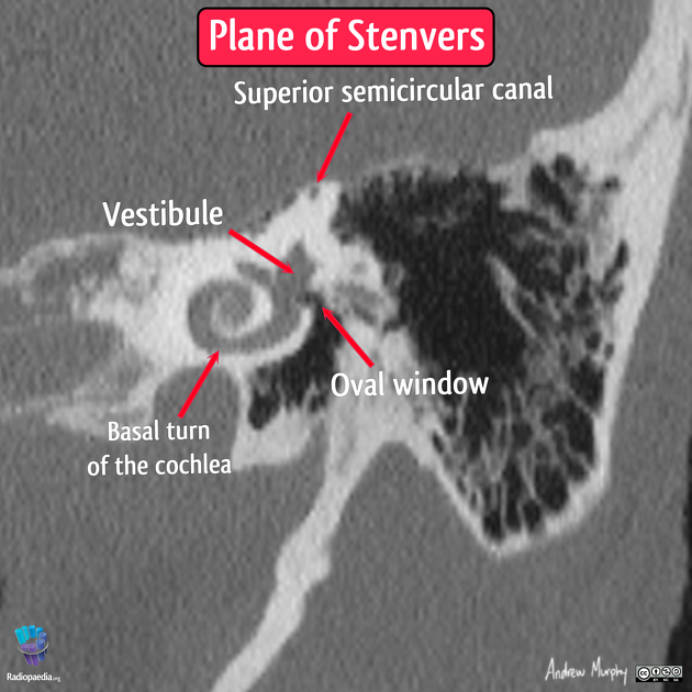
In addition to axial, coronal and sagittal views, oblique views along the long axis the bone (Stenvers view) or at right angles to this (Pöschl projection) are useful.


 Unable to process the form. Check for errors and try again.