
Sphenoethmoidal air cell
Citation, DOI, disclosures and article data
At the time the article was created Frank Gaillard had no recorded disclosures.
View Frank Gaillard's current disclosuresAt the time the article was last revised Bence Paládi had no financial relationships to ineligible companies to disclose.
View Bence Paládi's current disclosures- Sphenoethmoidal air cells
- Onodi cell
- Sphenoethmoidal air cell
- Central Onodi cell
- Onodi air cell
- Central Onodi air cell
- Onodi air cells
- Onodi cells
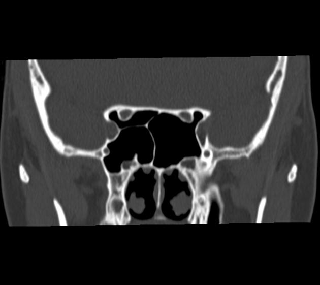
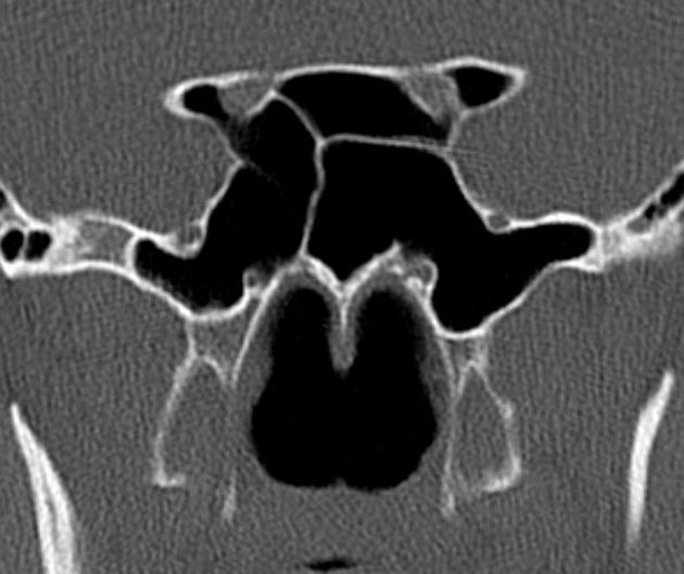
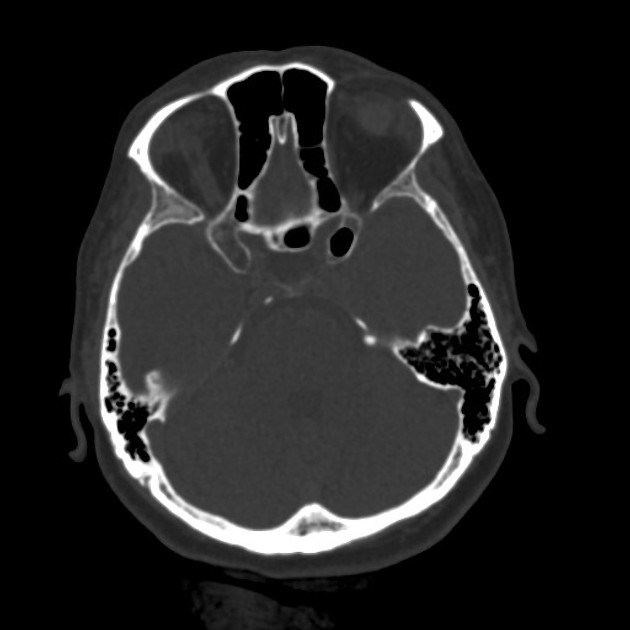
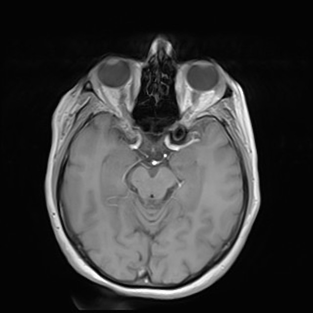
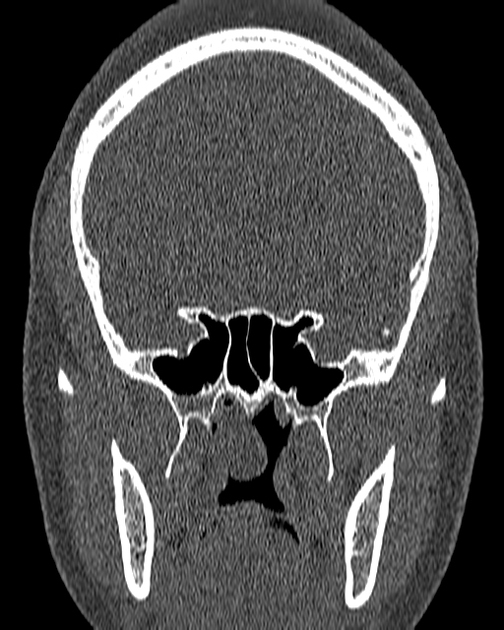
Sphenoethmoidal air cell, also commonly known as the Onodi air cell, is an anatomical variant of the paranasal sinuses, important due to its close proximity to the optic nerve and internal carotid artery.
On this page:
Terminology
The sphenoethmoidal air cell is generally defined as the most posterior ethmoidal air cell, that extends posteriorly to lie superolateral to the sphenoid sinus and thus in close proximity to the optic nerve and internal carotid artery 5,8. It often extends into the anterior clinoid process; importantly, aeration of the anterior clinoid process does not imply presence of an Onodi cell, as frequently such aeration is due to rescesses of the sphenoid sinus 9.
Rarely, it may lie superiorly to the sphenoid sinus and is then called a central Onodi air cell 4.
Epidemiology
The incidence of sphenoethmoidal air cells is variable, reported in 3.4 to 60% of individuals 1,8; the discrepancy of these rates of incidence likely results from variability in definition rather than true incidence 8.
Clinical presentation
Onodi air cells are usually asymptomatic, unless complicated by sinus disease (e.g. mucocoeles, squamous cell carcinoma, and acute sinusitis, which have all been reported in sphenoethmoidal air cells), leading to early optic nerve involvement.
The importance of these air cells stems primarily from their location adjacent to the optic nerve and internal carotid artery, with as little as 0.03 mm (median 0.08 mm) of bone separating them 1.
Potential damage to these critical structures occurs when attempts to enter the sphenoid sinus endoscopically are made by passing through the posterior wall of the sphenoethmoidal air cell expecting to enter the sphenoid sinus 5.
History and etymology
The importance and variability of the posteriormost ethmoidal air cells was described by the Hungarian laryngologist Adolf Ónodi (1857-1920) in 1904 6,7.
See also
concha bullosa: aerated middle turbinate
agger nasi cells: anteriormost ethmoidal cells
Haller cells: infraorbital ethmoidal air cells
Quiz questions
References
- 1. Thanaviratananich S, Chaisiwamongkol K, Kraitrakul S et-al. The prevalence of an Onodi cell in adult Thai cadavers. Ear Nose Throat J. 2003;82 (3): 200-4. - Pubmed citation
- 2. Lim CC, Dillon WP, Mcdermott MW. Mucocele involving the anterior clinoid process: MR and CT findings. AJNR Am J Neuroradiol. 1999;20 (2): 287-90. AJNR Am J Neuroradiol (full text) - Pubmed citation
- 3. Jones NS, Strobl A, Holland I. A study of the CT findings in 100 patients with rhinosinusitis and 100 controls. Clin Otolaryngol Allied Sci. 1997;22 (1): 47-51. - Pubmed citation
- 4. Cherla DV, Tomovic S, Liu JK et-al. The central Onodi cell: A previously unreported anatomic variation. Allergy Rhinol (Providence). 2013;4 (1): e49-51. doi:10.2500/ar.2013.4.0047 - Free text at pubmed - Pubmed citation
- 5. Kennedy DW, Bolger WE, Zinreich SJ. Diseases of the Sinuses. pmph usa. (2001) ISBN:1550090453. Read it at Google Books - Find it at Amazon
- 6. Onodi A, Die Sehstörungen und Erblindung nasalen Ursprunges, bedingt durch Erkrankungen der hinteren Nebenhöhlen. Ophthalmologica 1904;12:23-46
- 7. Forbis PTL. Stedman's Medical Eponyms. Lippincott Williams & Wilkins. (2005) ISBN:0781754437. Read it at Google Books - Find it at Amazon
- 8. Matti Anniko, Manuel Bernal-Sprekelsen, Victor Bonkowsky, Patrick Bradley, Salvatore Iurato. Otorhinolaryngology, Head and Neck Surgery. Springer (2010). ISBN:3540689400. Read it at Google Books - Find it at Amazon
- 9. Lim CC, Dillon WP, McDermott MW. Mucocele involving the anterior clinoid process: MR and CT findings. AJNR Am J Neuroradiol. 1999;20 (2): 287-90. AJNR Am J Neuroradiol (full text) - Pubmed citation
- 10. William T. O’Brien, Sr, Stefan Hamelin, Erik K. Weitzel. The Preoperative Sinus CT: Avoiding a “CLOSE” Call with Surgical Complications. (2016) Radiology. 281 (1): 10-21. doi:10.1148/radiol.2016152230 - Pubmed
Incoming Links
Related articles: Anatomy: Head and neck
- skeleton of the head and neck
-
cranial vault
- scalp (mnemonic)
- fontanelle
-
sutures
- calvarial
- facial
- frontozygomatic suture
- frontomaxillary suture
- frontolacrimal suture
- frontonasal suture
- temporozygomatic suture
- zygomaticomaxillary suture
- parietotemporal suture (parietomastoid suture)
- occipitotemporal suture (occipitomastoid suture)
- sphenofrontal suture
- sphenozygomatic suture
- spheno-occipital suture (not a true suture)
- lacrimomaxillary suture
- nasomaxillary suture
- internasal suture
- basal/internal
- skull landmarks
- frontal bone
- temporal bone
- parietal bone
- occipital bone
- skull base (foramina)
-
facial bones
- midline single bones
- paired bilateral bones
- cervical spine
- hyoid bone
- laryngeal cartilages
-
cranial vault
- muscles of the head and neck
- muscles of the tongue (mnemonic)
- muscles of mastication
-
facial muscles
- epicranius muscle
- circumorbital and palpebral muscles
- nasal muscles
-
buccolabial muscles
- elevators, retractors and evertors of the upper lip
- levator labii superioris alaeque nasalis muscle
- levator labii superioris muscle
- zygomaticus major muscle
- zygomaticus minor muscle
- levator anguli oris muscle
- malaris muscle
- risorius muscle
- depressors, retractors and evertors of the lower lip
- depressor labii inferioris muscle
- depressor anguli oris muscle
- mentalis muscle
- compound sphincter
-
orbicularis oris muscle
- incisivus labii superioris muscle
- incisivus labii inferioris muscle
-
orbicularis oris muscle
- muscle of mastication
- modiolus
- elevators, retractors and evertors of the upper lip
- muscles of the middle ear
- orbital muscles
- muscles of the soft palate
- pharyngeal muscles
- suprahyoid muscles
- infrahyoid muscles
- intrinsic muscles of the larynx
- muscles of the neck
- platysma muscle
- longus colli muscle
- longus capitis muscle
- scalenus anterior muscle
- scalenus medius muscle
- scalenus posterior muscle
- scalenus pleuralis muscle
- sternocleidomastoid muscle
-
suboccipital muscles
- rectus capitis posterior major muscle
- rectus capitis posterior minor muscle
- obliquus capitis superior muscle
- obliquus capitis inferior muscle
- accessory muscles of the neck
- deep cervical fascia
-
deep spaces of the neck
- anterior cervical space
- buccal space
- carotid space
- danger space
- deep cervical fascia
- infratemporal fossa
- masticator space
- parapharyngeal space
- stylomandibular tunnel
- parotid space
- pharyngeal (superficial) mucosal space
- perivertebral space
- posterior cervical space
- pterygopalatine fossa
- retropharyngeal space
- suprasternal space (of Burns)
- visceral space
- surgical triangles of the neck
- orbit
- ear
- paranasal sinuses
- upper respiratory tract
- viscera of the neck
- blood supply of the head and neck
-
arterial supply
-
common carotid artery
- carotid body
- carotid bifurcation
- subclavian artery
- variants
-
common carotid artery
- venous drainage
-
arterial supply
- innervation of the head and neck
-
cranial nerves
- olfactory nerve (CN I)
- optic nerve (CN II)
- oculomotor nerve (CN III)
- trochlear nerve (CN IV)
-
trigeminal nerve (CN V) (mnemonic)
- trigeminal ganglion
- ophthalmic division
- maxillary division
- mandibular division
- abducens nerve (CN VI)
- facial nerve (CN VII)
-
vestibulocochlear nerve (CN VIII)
- vestibular ganglion (Scarpa's ganglion)
- glossopharyngeal nerve (CN IX)
- vagus nerve (CN X)
- (spinal) accessory nerve (CN XI)
- hypoglossal nerve (CN XII)
- parasympathetic ganglia of the head and neck
- cervical sympathetic ganglia
- greater occipital nerve
- third occipital nerve
-
cervical plexus
- muscular branches
- longus capitis
- longus colli
- scalenes
- geniohyoid
- thyrohyoid
-
ansa cervicalis
- omohyoid (superior and inferior bellies separately)
- sternothyroid
- sternohyoid
- phrenic nerve
- contribution to the accessory nerve (CN XI)
- cutaneous branches
- muscular branches
- brachial plexus
- pharyngeal plexus
-
cranial nerves
- lymphatic drainage of the head and neck
- embryological development of the head and neck


 Unable to process the form. Check for errors and try again.