
Sternothyroid muscle
Citation, DOI, disclosures and article data
At the time the article was created Chamath Ariyasinghe had no recorded disclosures.
View Chamath Ariyasinghe's current disclosuresAt the time the article was last revised Rose Mark had no recorded disclosures.
View Rose Mark's current disclosures- Sternothyroid muscles
- Sterno-thyroid muscle
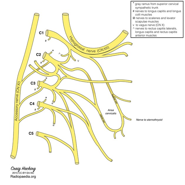
The sternothyroid muscle is an infrahyoid muscle of the neck that is innervated by the ansa cervicalis of the cervical plexus receiving fibers from the ventral rami of C1-C3 spinal nerves. The sternothyroid is a paired, flat strap of muscle that serves to fix the hyoid bone as well as depressing the larynx in phonation and in the terminal phase of swallowing.
On this page:
Summary
- origin: posterior surface of manubrium sterni
- insertion: oblique line on the lamina of the thyroid cartilage
- innervation: anterior rami of C1-C3 spinal nerves through the ansa cervicalis
- action: depresses and fixes the hyoid bone
Gross anatomy
Origin
The sternothyroid muscles are paired broad, flat muscles that lie deep to the sternohyoid muscles. They originate from the lower part of the posterior surface of the manubrium so far inferiorly that there is no attachment to the clavicle or sternoclavicular joints.
Insertion
The muscle travels vertically upwards to attach to the oblique line on the lamina of the thyroid cartilage end-to-end with the thyrohyoid muscle.
Relations
The sternothyroid muscle is one of the infrahyoid “strap” muscles and is not easily visualized as it lies deep to the sternohyoid muscle. At its origin the sternothyroid muscle extends as far laterally as the first costal cartilage and as it ascends into the neck it covers the lateral lobe of the thyroid gland.
Blood supply
The sternothyroid muscles receive their blood supply from the superior thyroid arteries.
Innervation
The sternothyroid muscles are innervated by the anterior rami of C1-C3 (predominantly C2 and C3) through the ansa cervicalis of the cervical plexus.
Action
The sternothyroid muscles primarily depress and fix the hyoid bone and underlying larynx.
References
- 1. Dalley AF, Agur AM. Clinically Oriented Anatomy, Sixth Edition. Lippincott Williams & Wilkins. ISBN:1605476528. Read it at Google Books - Find it at Amazon
- 2. Schuenke M, Schulte E, Schumacher U et-al. Neck and Internal Organs. Thieme. ISBN:1604062886. Read it at Google Books - Find it at Amazon
- 3. Lasts Anatomy Regional and Applied. CHURCHILL LIVINGSTONE. (2003) ISBN:B0084AQDG8. Read it at Google Books - Find it at Amazon
- 4. Kademani D, Tiwana P. Atlas of Oral and Maxillofacial Surgery, 1e. Saunders. ISBN:1455753289. Read it at Google Books - Find it at Amazon
Incoming Links
- Manubrium
- Superior mediastinum
- Brachiocephalic vein
- Ansa cervicalis
- Sternohyoid muscle
- Cervical plexus
- Thyroid cartilage
- Middle layer of the deep cervical fascia
- Differentiated thyroid cancer (staging)
- Infrahyoid muscles
- Muscular triangle
- Sternoclavicular joint
- Medullary thyroid cancer (staging)
- Middle thyroid vein
- Superior thoracic aperture
- Extrinsic muscles of the larynx
- Sternum
- Anaplastic thyroid cancer (staging)
Related articles: Anatomy: Head and neck
- skeleton of the head and neck
-
cranial vault
- scalp (mnemonic)
- fontanelle
-
sutures
- calvarial
- facial
- frontozygomatic suture
- frontomaxillary suture
- frontolacrimal suture
- frontonasal suture
- temporozygomatic suture
- zygomaticomaxillary suture
- parietotemporal suture (parietomastoid suture)
- occipitotemporal suture (occipitomastoid suture)
- sphenofrontal suture
- sphenozygomatic suture
- spheno-occipital suture (not a true suture)
- lacrimomaxillary suture
- nasomaxillary suture
- internasal suture
- basal/internal
- skull landmarks
- frontal bone
- temporal bone
- parietal bone
- occipital bone
- skull base (foramina)
-
facial bones
- midline single bones
- paired bilateral bones
- cervical spine
- hyoid bone
- laryngeal cartilages
-
cranial vault
- muscles of the head and neck
- muscles of the tongue (mnemonic)
- muscles of mastication
-
facial muscles
- epicranius muscle
- circumorbital and palpebral muscles
- nasal muscles
-
buccolabial muscles
- elevators, retractors and evertors of the upper lip
- levator labii superioris alaeque nasalis muscle
- levator labii superioris muscle
- zygomaticus major muscle
- zygomaticus minor muscle
- levator anguli oris muscle
- malaris muscle
- risorius muscle
- depressors, retractors and evertors of the lower lip
- depressor labii inferioris muscle
- depressor anguli oris muscle
- mentalis muscle
- compound sphincter
-
orbicularis oris muscle
- incisivus labii superioris muscle
- incisivus labii inferioris muscle
-
orbicularis oris muscle
- muscle of mastication
- modiolus
- elevators, retractors and evertors of the upper lip
- muscles of the middle ear
- orbital muscles
- muscles of the soft palate
- pharyngeal muscles
- suprahyoid muscles
- infrahyoid muscles
- intrinsic muscles of the larynx
- muscles of the neck
- platysma muscle
- longus colli muscle
- longus capitis muscle
- scalenus anterior muscle
- scalenus medius muscle
- scalenus posterior muscle
- scalenus pleuralis muscle
- sternocleidomastoid muscle
-
suboccipital muscles
- rectus capitis posterior major muscle
- rectus capitis posterior minor muscle
- obliquus capitis superior muscle
- obliquus capitis inferior muscle
- accessory muscles of the neck
- deep cervical fascia
-
deep spaces of the neck
- anterior cervical space
- buccal space
- carotid space
- danger space
- deep cervical fascia
- infratemporal fossa
- masticator space
- parapharyngeal space
- stylomandibular tunnel
- parotid space
- pharyngeal (superficial) mucosal space
- perivertebral space
- posterior cervical space
- pterygopalatine fossa
- retropharyngeal space
- suprasternal space (of Burns)
- visceral space
- surgical triangles of the neck
- orbit
- ear
- paranasal sinuses
- upper respiratory tract
- viscera of the neck
- blood supply of the head and neck
-
arterial supply
-
common carotid artery
- carotid body
- carotid bifurcation
- subclavian artery
- variants
-
common carotid artery
- venous drainage
-
arterial supply
- innervation of the head and neck
-
cranial nerves
- olfactory nerve (CN I)
- optic nerve (CN II)
- oculomotor nerve (CN III)
- trochlear nerve (CN IV)
-
trigeminal nerve (CN V) (mnemonic)
- trigeminal ganglion
- ophthalmic division
- maxillary division
- mandibular division
- abducens nerve (CN VI)
- facial nerve (CN VII)
-
vestibulocochlear nerve (CN VIII)
- vestibular ganglion (Scarpa's ganglion)
- glossopharyngeal nerve (CN IX)
- vagus nerve (CN X)
- (spinal) accessory nerve (CN XI)
- hypoglossal nerve (CN XII)
- parasympathetic ganglia of the head and neck
- cervical sympathetic ganglia
- greater occipital nerve
- third occipital nerve
-
cervical plexus
- muscular branches
- longus capitis
- longus colli
- scalenes
- geniohyoid
- thyrohyoid
-
ansa cervicalis
- omohyoid (superior and inferior bellies separately)
- sternothyroid
- sternohyoid
- phrenic nerve
- contribution to the accessory nerve (CN XI)
- cutaneous branches
- muscular branches
- brachial plexus
- pharyngeal plexus
-
cranial nerves
- lymphatic drainage of the head and neck
- embryological development of the head and neck


 Unable to process the form. Check for errors and try again.