
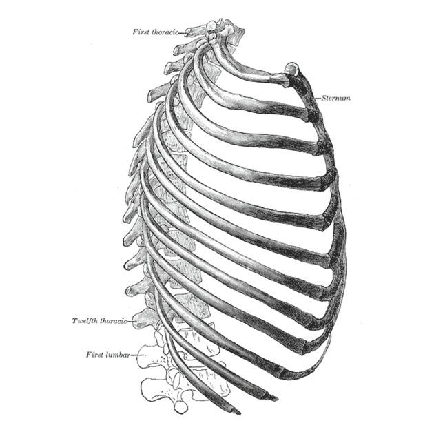
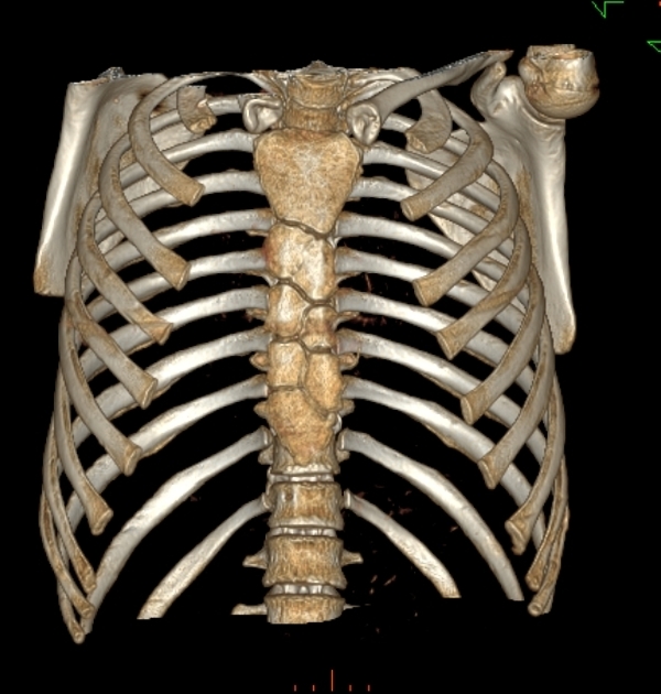
The sternum (plural: sterna or sternums) is the anterior midline chest wall bone plate that articulates with clavicles and ribs. It is composed of 3 parts (from superior to inferior): the manubrium, sternal body and xiphoid process.
On this page:
Gross anatomy
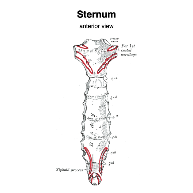
The sternum comprises three bones which are firmly connected by symphyses (secondary cartilaginous joints).
The broader superior surface of the quadrilateral manubrium has a midline notch, the sternal notch. The manubrium is connected to the deep cervical fascia, three neck muscles and is notched at the sternoclavicular and first costochondral joints. See manubrium for more detail.
The body is a flat rectangular bone approximately 20 cm long, 3-4 cm wide and 1 cm thick that has concavities (facets) on its lateral border at the rib articulations. See sternal body for more detail.
The xiphoid process is a thin bony projection inferiorly. See xiphoid process for more detail.
Articulations
-
the manubrium articulates with the:
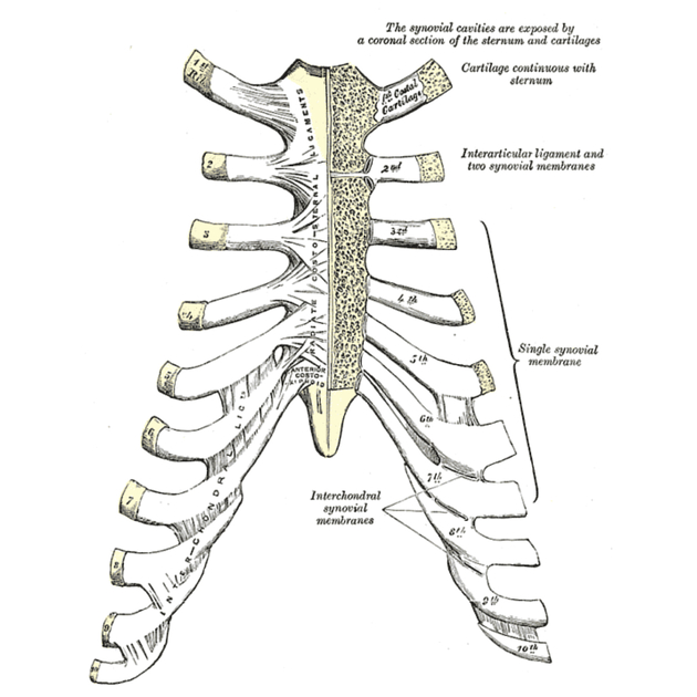
costal cartilages of the first rib via the sternocostal joints which are primary cartilaginous/synchondrosis joints,
clavicles via the sternoclavicular joints which are atypical synovial joints
body of the sternum via the manubriosternal joint which is a secondary cartilaginous/symphysis joint typically 6
-
the body articulates with the
costal cartilage of the second rib at the sternal angle (of Louis, which is in the transthoracic plane of Ludwig) and the
-
costal cartilages of the 3rd to 7th ribs
articulation of 2nd costal cartilages are shared as demifacets between the manubrium and the sternal body
articulations 7th costal cartilages are shared as demifacets between the sternal body and the xiphoid, respectively
xiphoid process via the xiphisternal joint which is a secondary cartilaginous/symphysis joint 6
Attachments
Musculotendinous
muscular attachments: sternocleidomastoid, intercostal, pectoralis major, sternohyoid, and sternothyroid muscles
transversus thoracis muscle arises from the posterior surface of the body
the xiphisternum attaches to linea alba
Ligamentous
sternopericardial ligaments secure the fibrous pericardium to it
interclavicular ligament
anterior and posterior sternoclavicular ligaments (thickenings of the sternoclavicular joint capsule)
Arterial supply
nutrient branches from internal thoracic (mammary) artery
deep to the sternum is the internal thoracic artery laterally
Venous drainage
tributaries of the internal thoracic veins, posterolateral to the sternum
Innervation
nerve supply is via intercostal nerves which arise from the anterior rami of thoracic spinal nerves
Lymphatic drainage
chain of internal mammary lymph nodes
Variant anatomy
tilted sternum: oblique instead of horizontal orientation in the mediolateral plane 5
unfused sternal body segments (sternebrae)
Development
Ossification
The manubrium forms from a single primary ossification center present at birth. The body of the sternum typically develops from four sternebrae (horizontal bony bars) with the first sternebra most often arising from a single primary ossification center prenatally and the second to fourth sternebrae most often arising from two or more primary ossification centers within each sternebra postnatally 8. Ossification is observed to occur in a superior to inferior direction, whilst fusion of the ossification centers between adjacent sternebrae occurs in an inferosuperior direction. Ossification continues up to 18-19 years of age with fusion commencing as early as 7 years in some individuals. The xiphisternum ossifies quite independently later in life.
A number of sternal variants arise due to morphological differences in ossification and fusion such as a sternal foramen, unfused sternebrae, or honeycomb sternum.


 Unable to process the form. Check for errors and try again.