
Testicular appendix
Citation, DOI, disclosures and article data
At the time the article was created Vasileios Rafailidis had no recorded disclosures.
View Vasileios Rafailidis's current disclosuresAt the time the article was last revised Tariq Walizai had no financial relationships to ineligible companies to disclose.
View Tariq Walizai's current disclosures- Appendix of testis
- Testicular appendix
- Αppendix of the testis
- Αppendix of testis
- Appendix testis
- Hydatid of Morgagni (testis)
A testicular appendix (alternatively called appendix of testis or appendix testis, and historically also known as hydatid of Morgagni) represents a developmental remnant of the paramesonephric duct (Müllerian duct) which is situated in the upper pole of the testis inside a groove between the testis and the head of epididymis 1.
On this page:
Epidemiology
The prevalence in childhood is around 85% (range 83-92%). The testicular appendix can be bilateral in 69% of the cases 4.
Clinical presentation
Testicular appendages in general alone have no clinical significance. However, when complicated by torsion, they can lead to acute scrotal pain 2.
Radiographic features
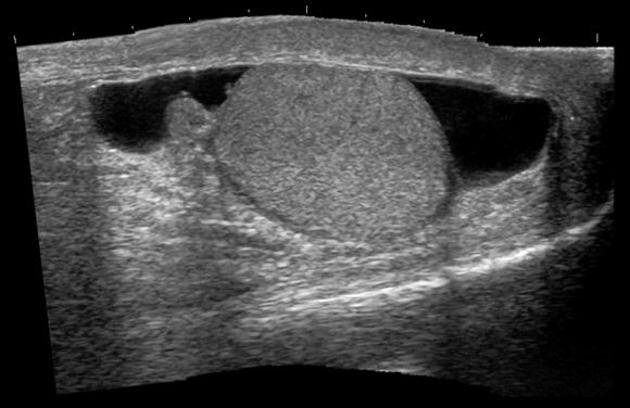
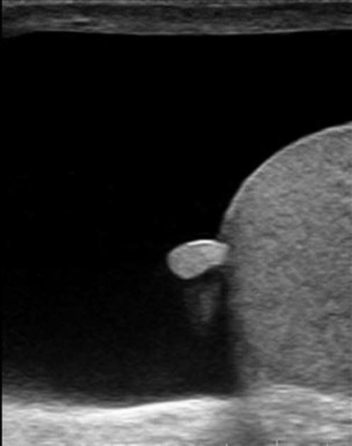
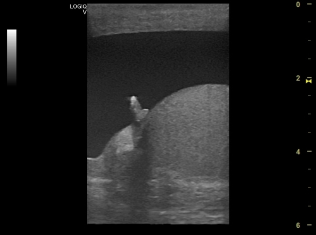
Ultrasound
Ultrasonography with high-frequency linear array transducers is the modality of choice in evaluating the scrotum and testis with its appendages. It can quickly and easily demonstrate the existence of such anatomic structures and their vascularity without exposure to radiation.
Appendages of the testis are best seen when combined with hydrocele. The frequency of the ultrasonographic identification of these anatomic structures is around 89% 3.
The normal testicular appendix is seen as an oval, sessile structure in 88% of cases 4. Its length ranges from 1 to 7 mm 1. Color Doppler ultrasonography can occasionally detect blood flow inside testicular appendages 4. When pedunculated, the appendix is in danger of torsion.
History and etymology
"Appendix testis" was first described by Giovanni Battista Morgagni (1682-1771) in his textbook 'De Sedibus et Causis Morborum per Anatomen Indagatis' (On the Seats and Causes of Diseases Investigated by Anatomy) in 1761 5.
Differential diagnosis
In cases of torsion, patients present with acute scrotal pain and imaging differential diagnosis includes:
Related pathology
References
- 1. Sellars M & Sidhu P. Ultrasound Appearances of the Testicular Appendages: Pictorial Review. Eur Radiol. 2003;13(1):127-35. doi:10.1007/s00330-002-1387-1 - Pubmed
- 2. Valentino M, Bertolotto M, Ruggirello M, Pavlica P, Barozzi L, Rossi C. Cystic Lesions and Scrotal Fluid Collections in Adults: Ultrasound Findings. J Ultrasound. 2011;14(4):208-15. doi:10.1016/j.jus.2011.10.008 - Pubmed
- 3. Johnson K & Dewbury K. Ultrasound Imaging of the Appendix Testis and Appendix Epididymis. Clin Radiol. 1996;51(5):335-7. doi:10.1016/s0009-9260(96)80110-3 - Pubmed
- 4. Baldisserotto M, de Souza J, Pertence A, Dora M. Color Doppler Sonography of Normal and Torsed Testicular Appendages in Children. AJR Am J Roentgenol. 2005;184(4):1287-92. doi:10.2214/ajr.184.4.01841287 - Pubmed
- 5. Zani A & Cozzi D. Giovanni Battista Morgagni and His Contribution to Pediatric Surgery. J Pediatr Surg. 2008;43(4):729-33. doi:10.1016/j.jpedsurg.2007.12.065 - Pubmed
Incoming Links
Related articles: Anatomy: Abdominopelvic
- skeleton of the abdomen and pelvis
- muscles of the abdomen and pelvis
- spaces of the abdomen and pelvis
- anterior abdominal wall
- posterior abdominal wall
- abdominal cavity
- pelvic cavity
- perineum
- abdominal and pelvic viscera
- gastrointestinal tract
- spleen
- hepatobiliary system
-
endocrine system
-
adrenal gland
- adrenal vessels
- chromaffin cells
- variants
- pancreas
- organs of Zuckerkandl
-
adrenal gland
-
urinary system
-
kidney
- renal pelvis
- renal sinus
- avascular plane of Brodel
-
variants
- number
- fusion
- location
- shape
- ureter
- urinary bladder
- urethra
- embryology
-
kidney
- male reproductive system
-
female reproductive system
- vulva
- vagina
- uterus
- adnexa
- Fallopian tubes
- ovaries
- broad ligament (mnemonic)
- variant anatomy
- embryology
- blood supply of the abdomen and pelvis
- arteries
-
abdominal aorta
- inferior phrenic artery
- celiac artery
- superior mesenteric artery
- middle suprarenal artery
- renal artery (variant anatomy)
- gonadal artery (ovarian artery | testicular artery)
- inferior mesenteric artery
- lumbar arteries
- median sacral artery
-
common iliac artery
- external iliac artery
-
internal iliac artery (mnemonic)
- anterior division
- umbilical artery
- superior vesical artery
- obturator artery
- vaginal artery
- inferior vesical artery
- uterine artery
- middle rectal artery
-
internal pudendal artery
- inferior rectal artery
-
perineal artery
- posterior scrotal artery
- transverse perineal artery
- artery to the bulb
- deep artery of the penis/clitoris
- dorsal artery of the penis/clitoris
- inferior gluteal artery
- posterior division (mnemonic)
- variant anatomy
- anterior division
-
abdominal aorta
- portal venous system
- veins
- anastomoses
- arterioarterial anastomoses
- portal-systemic venous collateral pathways
- watershed areas
- arteries
- lymphatics
- innervation of the abdomen and pelvis
- thoracic splanchnic nerves
- lumbar plexus
-
sacral plexus
- lumbosacral trunk
- sciatic nerve
- superior gluteal nerve
- inferior gluteal nerve
- nerve to piriformis
- perforating cutaneous nerve
- posterior femoral cutaneous nerve
- parasympathetic pelvic splanchnic nerves
- pudendal nerve
- nerve to quadratus femoris and inferior gemellus muscles
- nerve to internal obturator and superior gemellus muscles
- autonomic ganglia and plexuses


 Unable to process the form. Check for errors and try again.