
Variant hepatic arterial anatomy
Citation, DOI, disclosures and article data
At the time the article was created Donna D'Souza had no recorded disclosures.
View Donna D'Souza's current disclosuresAt the time the article was last revised gaia russo had no financial relationships to ineligible companies to disclose.
View gaia russo's current disclosures- Variantion in hepatic arterial anatomy
- Hepatic arterial variations
- Hepatic arterial variants
- Variation of the hepatic artery
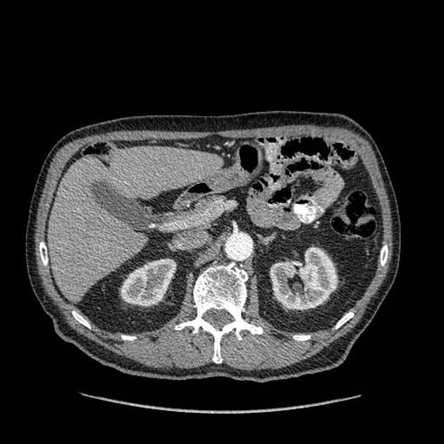
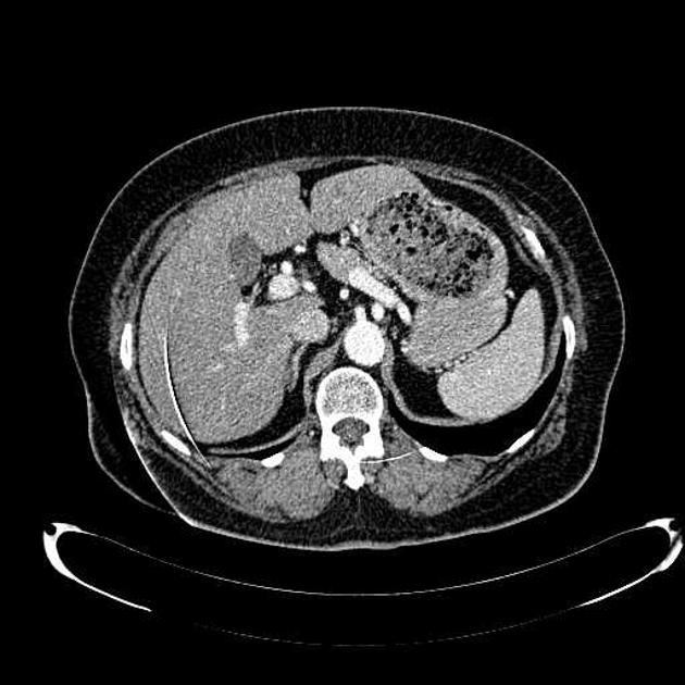
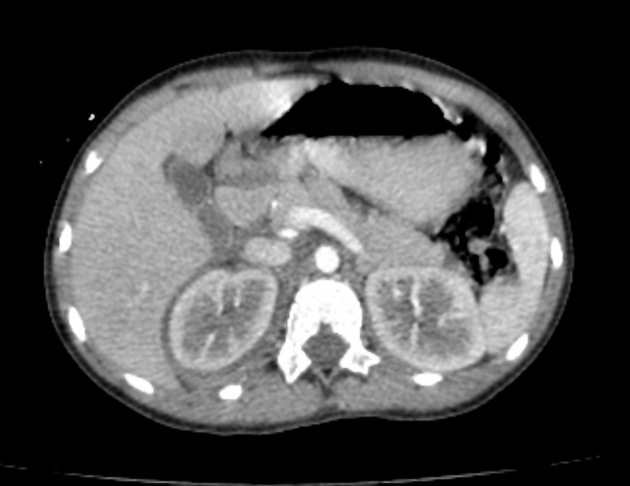
Variation in hepatic arterial anatomy is seen in 40-45% of people. Classic branching of the common hepatic artery from the celiac artery, and the proper hepatic artery into right and left hepatic arteries to supply the entire liver, is seen in 55-60% of the population.
On this page:
Terminology
An accessory hepatic artery is one which arises from an anomalous origin and supplies a portion of the liver along with another artery.
A replaced hepatic artery is one which arises from an anomalous origin and supplies a portion of the liver solely.
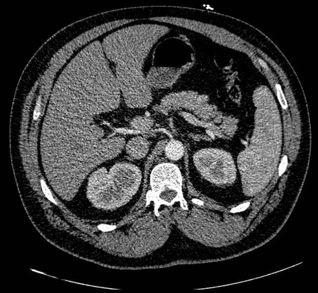
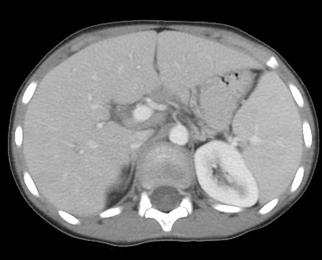
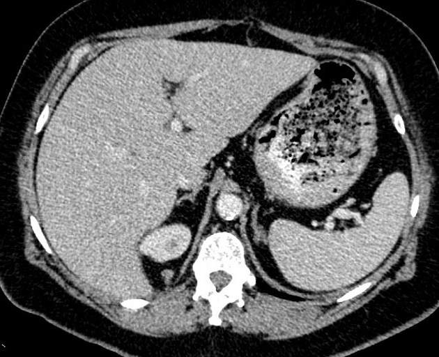
Variant anatomy
In general, the common hepatic artery may arise from the abdominal aorta or superior mesenteric artery (SMA) and all or part of the right and left hepatic arteries may arise from (be replaced to) other vessels.
The two most common variants are:
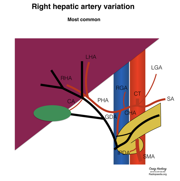
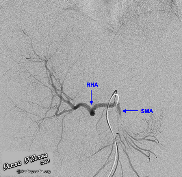
replaced right hepatic artery arising from the SMA
replaced left hepatic artery arising from the left gastric artery
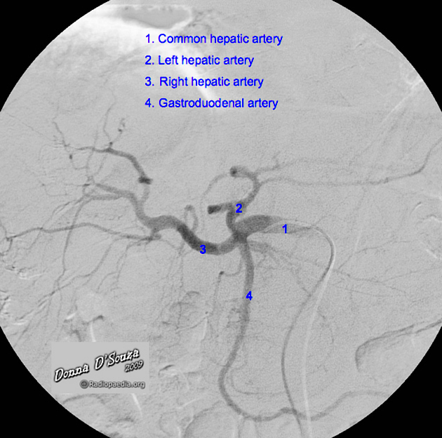
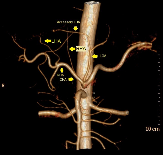
Another common finding, though not considered a variant by many authors, is trifurcation of the common hepatic artery into the right hepatic artery, left hepatic artery and gastroduodenal artery (GDA). With this branching pattern, there is no proper hepatic artery (PHA).
Common hepatic artery (CHA)
from aorta: 2%
from SMA: 2%
trifurcation into RHA, LHA and GDA: ~6% (range 4-8%)
Right hepatic artery (RHA)
from celiac artery: ~2.5% (range 1-4%)
from SMA: ~12.5% (range 9-15%) 7
accessory right hepatic artery from SMA: ~4% (range 1-7%) 7
Left hepatic artery (LHA)
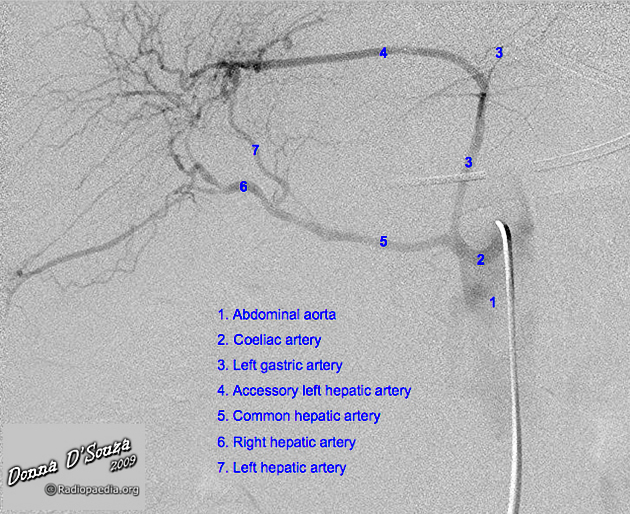
from left gastric artery (LGA): ~7.5% (range 4-11%) 7
accessory left hepatic artery from LGA: ~7.5% (range 4-11%)
Right and left hepatic arteries
RHA from SMA and LHA from LGA: ~1% (range 0.5-2%)
accessory RHA and LHA: 1%
Middle hepatic artery (MHA)
middle hepatic artery usually arises from the left hepatic artery
it may arise from the right hepatic artery
it may arise as a trifurcation of the proper hepatic artery in the porta hepatis
in patients with a replaced left hepatic artery, the middle hepatic artery arises from the right hepatic artery
in patients with a replaced right hepatic artery, the middle hepatic artery arises from the left hepatic artery
if the bifurcation of the proper hepatic artery is low within the porta hepatis, then the middle hepatic artery may have an extrahepatic course and traverse Calot's triangle
Classification
A classification method was described by Michel et al. in 1955 6:
I: standard anatomy ~60% (range 55-61%)
II: replaced LHA ~7.5% (range 3-10%)
III: replaced RHA ~10% (range 8-11 %)
IV: replaced RHA and LHA ~1%
V: accessory LHA from LGA ~10% (range 8-11%)
VI: accessory RHA from SMA ~5% (range 1.5-7%)
VII: accessory RHA and LHA ~1%
VIII: accessory RHA and LHA and replaced LHA or RHA ~2.5%
IX: CHA replaced to SMA ~3% (range 2-4.5%)
X: CHA replaced to LGA ~0.5%
-
unclassified
CHA separate origin from aorta ~2%
double hepatic artery ~4%
PHA replaced to SMA; GDA origin from aorta <0.5%
See also
References
- 1. Kaufman JA, Lee MJ. Vascular & interventional radiology. Mosby Inc. (2004) ISBN:0815143699. Read it at Google Books - Find it at Amazon
- 2. Covey AM, Brody LA, Maluccio MA et-al. Variant hepatic arterial anatomy revisited: digital subtraction angiography performed in 600 patients. Radiology. 2002;224 (2): 542-7. doi:10.1148/radiol.2242011283 - Pubmed citation
- 3. Winston CB, Lee NA, Jarnagin WR et-al. CT angiography for delineation of celiac and superior mesenteric artery variants in patients undergoing hepatobiliary and pancreatic surgery. AJR Am J Roentgenol. 2007;189 (1): W13-9. doi:10.2214/AJR.04.1374 - Pubmed citation
- 4. Song SY, Chung JW, Yin YH et-al. Celiac axis and common hepatic artery variations in 5002 patients: systematic analysis with spiral CT and DSA. Radiology. 2010;255 (1): 278-88. doi:10.1148/radiol.09090389 - Pubmed citation
- 5. Ugurel MS, Battal B, Bozlar U et-al. Anatomical variations of hepatic arterial system, coeliac trunk and renal arteries: an analysis with multidetector CT angiography. Br J Radiol. 2010;83 (992): 661-7. doi:10.1259/bjr/21236482 - Pubmed citation
- 6. Covey AM, Brody LA, Maluccio MA et-al. Variant hepatic arterial anatomy revisited: digital subtraction angiography performed in 600 patients. Radiology. 2002;224 (2): 542-7. doi:10.1148/radiol.2242011283 - Pubmed citation
- 7. Stephanie Ryan, Michelle McNicholas, Stephen J. Eustace. Anatomy for Diagnostic Imaging. (2011) Page 178. ISBN: 9780702029714 - Google Books
Incoming Links
- Chemoembolization of hepatocellular carcinoma through an accessory right hepatic artery
- Antiphospholipid syndrome with adrenal insufficiency
- Superior mesenteric artery and replaced right hepatic artery thrombosis
- Hepatic hemangioma
- Calyceal diverticulum
- Common hepatic artery originating from aorta
- Oppenheimer ossicle
- Traumatic renal injury - AAST grade IV injury
- Isolated celiac and splenic artery dissection
- Coeliac trunk compression syndrome and aberrant left hepatic artery
- Circumaortic left renal vein with a duplicated retroaortic vein
- Accessory left hepatic artery
- Common hepatic artery anatomic variation (diagram)
- Right hepatic artery anatomic variation (diagram)
- Left hepatic artery anatomic variation (diagram)
- Hepatic artery pseudoaneurysm rupture
- Transplant hepatic artery - patch arterial anastomosis
- Replaced left and right hepatic arteries
- Replaced left and right hepatic arteries
- Congenital extrahepatic portosystemic shunt
Related articles: Anatomy: Abdominopelvic
- skeleton of the abdomen and pelvis
- muscles of the abdomen and pelvis
- spaces of the abdomen and pelvis
- anterior abdominal wall
- posterior abdominal wall
- abdominal cavity
- pelvic cavity
- perineum
- abdominal and pelvic viscera
- gastrointestinal tract
- spleen
- hepatobiliary system
-
endocrine system
-
adrenal gland
- adrenal vessels
- chromaffin cells
- variants
- pancreas
- organs of Zuckerkandl
-
adrenal gland
-
urinary system
-
kidney
- renal pelvis
- renal sinus
- avascular plane of Brodel
-
variants
- number
- fusion
- location
- shape
- ureter
- urinary bladder
- urethra
- embryology
-
kidney
- male reproductive system
-
female reproductive system
- vulva
- vagina
- uterus
- adnexa
- Fallopian tubes
- ovaries
- broad ligament (mnemonic)
- variant anatomy
- embryology
- blood supply of the abdomen and pelvis
- arteries
-
abdominal aorta
- inferior phrenic artery
- celiac artery
- superior mesenteric artery
- middle suprarenal artery
- renal artery (variant anatomy)
- gonadal artery (ovarian artery | testicular artery)
- inferior mesenteric artery
- lumbar arteries
- median sacral artery
-
common iliac artery
- external iliac artery
-
internal iliac artery (mnemonic)
- anterior division
- umbilical artery
- superior vesical artery
- obturator artery
- vaginal artery
- inferior vesical artery
- uterine artery
- middle rectal artery
-
internal pudendal artery
- inferior rectal artery
-
perineal artery
- posterior scrotal artery
- transverse perineal artery
- artery to the bulb
- deep artery of the penis/clitoris
- dorsal artery of the penis/clitoris
- inferior gluteal artery
- posterior division (mnemonic)
- variant anatomy
- anterior division
-
abdominal aorta
- portal venous system
- veins
- anastomoses
- arterioarterial anastomoses
- portal-systemic venous collateral pathways
- watershed areas
- arteries
- lymphatics
- innervation of the abdomen and pelvis
- thoracic splanchnic nerves
- lumbar plexus
-
sacral plexus
- lumbosacral trunk
- sciatic nerve
- superior gluteal nerve
- inferior gluteal nerve
- nerve to piriformis
- perforating cutaneous nerve
- posterior femoral cutaneous nerve
- parasympathetic pelvic splanchnic nerves
- pudendal nerve
- nerve to quadratus femoris and inferior gemellus muscles
- nerve to internal obturator and superior gemellus muscles
- autonomic ganglia and plexuses
Related articles: Pathology: Hepato-Pancreato-Biliary
- liver
- depositional disorders
- infection and inflammation
- liver abscess
- hepatic hydatid infection
- cirrhosis
- hepatitis
- cholecystitis
- cholangitis
- malignancy
- liver and intrahepatic bile duct tumors
- benign epithelial tumors
- hepatocellular hyperplasia
- hepatocellular adenoma
- hepatic/biliary cysts
- benign nonepithelial tumors
- primary malignant epithelial tumors
- hepatocellular carcinoma
- hepatocellular carcinoma variants
-
cholangiocarcinoma
- intra-hepatic
- mass-forming type
- periductal infiltrating type - Klatskin tumors
- intraductal growing type
- extra-hepatic/large duct type
- intra-hepatic
- biliary cystadenocarcinoma
- combined hepatocellular and cholangiocarcinoma
- hepatoblastoma
- undifferentiated carcinoma
- primary malignant nonepithelial tumors
- hematopoietic and lymphoid tumors
- primary hepatic lymphoma
- hepatic myeloid sarcoma (hepatic chloroma)
- secondary tumors
- miscellaneous
- adrenal rest tumors
- hepatic carcinosarcoma
- hepatic fibroma
- hepatic hemangioma
- hepatic Kaposi sarcoma
- hepatic lipoma
- hepatic mesenchymal hamartoma
- hepatic myxoma
- hepatic rhabdoid tumor
- hepatic solitary fibrous tumor
- hepatic teratoma
- hepatic yolk sac tumor
- inflammatory myofibroblastic tumor (inflammatory pseudotumor)
- nodular regenerative hyperplasia
- pancreatic rest tumors
- primary hepatic carcinoid
- benign epithelial tumors
- liver and intrahepatic bile duct tumors
- metabolic
- trauma
-
vascular and perfusion disorders
- portal vein related
- hepatic artery related
- hepatic veins related
- inferior vena cava related
- other
- third inflow
- liver thrombotic angiitis
- infra diaphragmatic total anomalous pulmonary venous return (TAPVR)
- hereditary hemorrhagic telangiectasia (Osler-Weber-Rendu disease)
- pancreas
-
pancreatic neoplasms
- cystic neoplasm (cystic pancreatic mass differential diagnosis)
- solid neoplasm
- non-epithelial pancreatic neoplasms
- others
- simple pancreatic cyst
-
pancreatitis (mnemonic for the causes)
- acute pancreatitis
- chronic pancreatitis
- Ascaris-induced pancreatitis
- tropical pancreatitis
- autoimmune pancreatitis
- emphysematous pancreatitis
- hypertriglyceridemia-induced pancreatitis
- hereditary pancreatitis
- pancreatitis associated with cystic fibrosis
- pancreaticopleural fistula
- segmental pancreatitis
- pancreatic atrophy
- pancreatic lipomatosis
- pancreatic trauma
- pancreatic transplant
-
pancreatic neoplasms
- gallbladder and biliary
- congenital malformations and anatomical variants
- gallstones
- gallbladder inflammation
- bile ducts inflammation
- gallbladder wall abnormalities
- other gallbladder abnormalities
- bile duct dilatation (differential)
- bile duct wall thickening (differential)
- bile ducts neoplasms


 Unable to process the form. Check for errors and try again.