
Cisterna chyli
Citation, DOI, disclosures and article data
At the time the article was created Yuranga Weerakkody had no recorded disclosures.
View Yuranga Weerakkody's current disclosuresAt the time the article was last revised Tariq Walizai had no financial relationships to ineligible companies to disclose.
View Tariq Walizai's current disclosures- Receptaculum chyli
- Pecquet's receptacle
- Pecquet receptacle
- Cisternae chyli
- Cisterna chyli (CC)
- Chyle cistern
The cisterna chyli (CC) (plural: cisternae chyli), also known as the receptaculum chyli, is a normal anatomical structure in the lymphatic system. It is seen as a saccular area of dilatation in the lymphatic channels that are located in the retrocrural space, usually to the immediate right of the origin of the abdominal aorta.
On this page:
Gross anatomy
Location
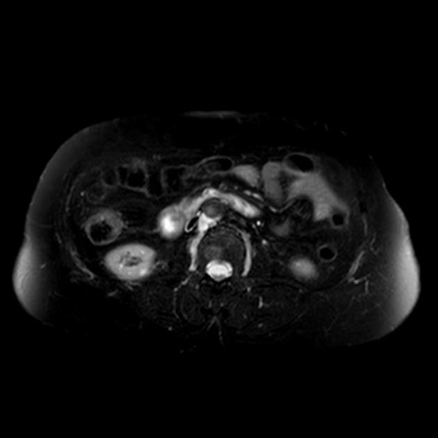
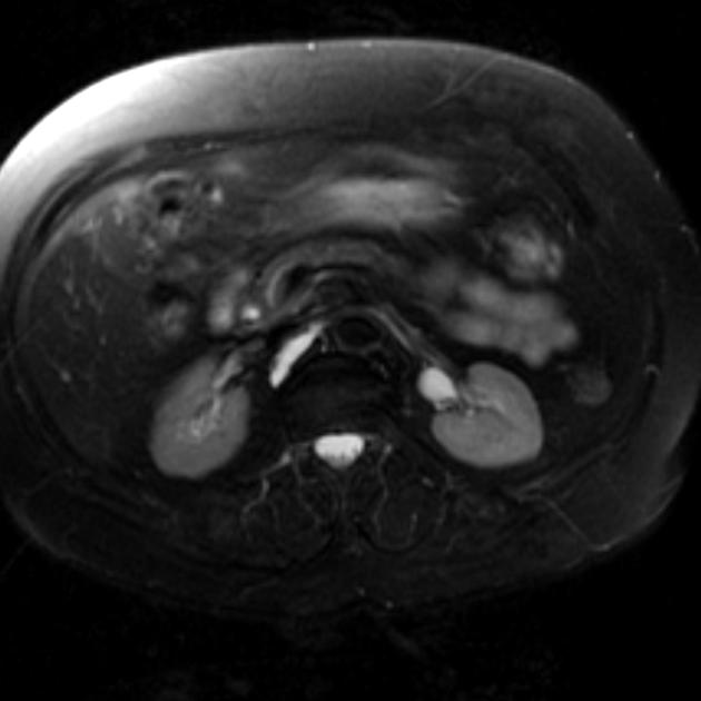
The cisterna chyli is located anterior to the L1 and L2 vertebral bodies, immediately right to the aorta behind the right crus of the diaphragm.
Origin
It is an elongated, sac-like structure formed by the junction of a variable number of lumbar, intestinal, liver and descending intercostal lymphatic trunks 7. It extends 5-7 cm in the caudocephalad axis, but the size varies greatly with patient position.
Termination
The upper end of the cisterna chyli continues as the thoracic duct which ascends in the posterior mediastinum to empty into the left subclavian vein.
Drainage
Receives lymph from the abdominal viscera as well as the abdominal wall (below the level of the umbilicus), non-alimentary viscera and lower extremities.
Relations
anterior: right crus of the diaphragm
posterior: L1 and L2 vertebral bodies
left lateral: abdominal aorta
right lateral: azygos vein
Radiographic features
CT
The cisterna chyli can be identified as a rounded-to-elliptical retrocrural structure with an average attenuation of 4 HU. There is no enhancement following intravenous contrast administration.
MRI
The signal intensity characteristics of the cisterna chyli on MRI are the same as those for static or slow-moving fluids with high signal intensity on fluid-sensitive MRI sequences 3.
History and etymology
Jean Pecquet (1624–1674), a French physician, in 1651, described in detail the thoracic duct and cisterna chyli, the latter being named Pecquet's receptacle, in his honor 6.
Differential diagnosis
On CT consider:
enlarged retrocrural lymph node 1
small neurenteric cyst
References
- 1. Gollub MJ, Castellino RA. The cisterna chyli: a potential mimic of retrocrural lymphadenopathy on CT scans. Radiology. 1996;199 (2): 477-80. Radiology (abstract) - Pubmed citation
- 2. Verma SK, Mitchell DG, Bergin D et-al. The cisterna chyli: enhancement on delayed phase MR images after intravenous administration of gadolinium chelate. Radiology. 2007;244 (3): 791-6. doi:10.1148/radiol.2443061518 - Pubmed citation
- 3. Erden A. Cisterna chyli: an incidental finding on MR cholangiopancreatography. AJR Am J Roentgenol. 2004;182 (1): 262. AJR Am J Roentgenol (full text) - Pubmed citation
- 4. Pinto PS, Sirlin CB, Andrade-barreto OA et-al. Cisterna chyli at routine abdominal MR imaging: a normal anatomic structure in the retrocrural space. Radiographics. 24 (3): 809-17. doi:10.1148/rg.243035086 - Pubmed citation
- 5. Smith TR, Grigoropoulos J. The cisterna chyli: incidence and characteristics on CT. Clin Imaging. 26 (1): 18-22. Clin Imaging (link) - Pubmed citation
- 6. Natale G, Bocci G, Ribatti D. Scholars and scientists in the history of the lymphatic system. (2017) Journal of anatomy. 231 (3): 417-429. doi:10.1111/joa.12644 - Pubmed
- 7. Kiyonaga M, Mori H, Matsumoto S, Yamada Y, Sai M, Okada F. Thoracic Duct and Cisterna Chyli: Evaluation with Multidetector Row CT. BJR. 2012;85(1016):1052-8. doi:10.1259/bjr/19379150 - Pubmed
Incoming Links
Related articles: Anatomy: General
- anatomic position
-
anatomic nomenclature
-
Terminologia Anatomica
- superseded nomenclature
-
Terminologia Anatomica
- anatomic variants
- labeled imaging anatomy cases
- regional anatomy
- systems anatomy
- endocrine system
- lymphatic system
- reticuloendothelial system
- nervous system
- systems based on location
- systems based on function
- somatic nervous system
-
autonomic nervous system
- sympathetic nervous system
- parasympathetic nervous system
-
autonomic ganglia and plexuses
- craniofacial
- cervical
- thoracic
- abdominopelvic
- coccygeal
- histology
- osteology
- skeleton
- bones
- macroscopic structure
- microscopic structure
- bone growth
- fetal bone formation
- developmental ossification
- tubulation
- bone types
- nutrient foramen
- joints
- muscles
- organs
- embryology
- skin
- blood vessels
Related articles: Anatomy: Abdominopelvic
- skeleton of the abdomen and pelvis
- muscles of the abdomen and pelvis
- spaces of the abdomen and pelvis
- anterior abdominal wall
- posterior abdominal wall
- abdominal cavity
- pelvic cavity
- perineum
- abdominal and pelvic viscera
- gastrointestinal tract
- spleen
- hepatobiliary system
-
endocrine system
-
adrenal gland
- adrenal vessels
- chromaffin cells
- variants
- pancreas
- organs of Zuckerkandl
-
adrenal gland
-
urinary system
-
kidney
- renal pelvis
- renal sinus
- avascular plane of Brodel
-
variants
- number
- fusion
- location
- shape
- ureter
- urinary bladder
- urethra
- embryology
-
kidney
- male reproductive system
-
female reproductive system
- vulva
- vagina
- uterus
- adnexa
- Fallopian tubes
- ovaries
- broad ligament (mnemonic)
- variant anatomy
- embryology
- blood supply of the abdomen and pelvis
- arteries
-
abdominal aorta
- inferior phrenic artery
- celiac artery
- superior mesenteric artery
- middle suprarenal artery
- renal artery (variant anatomy)
- gonadal artery (ovarian artery | testicular artery)
- inferior mesenteric artery
- lumbar arteries
- median sacral artery
-
common iliac artery
- external iliac artery
-
internal iliac artery (mnemonic)
- anterior division
- umbilical artery
- superior vesical artery
- obturator artery
- vaginal artery
- inferior vesical artery
- uterine artery
- middle rectal artery
-
internal pudendal artery
- inferior rectal artery
-
perineal artery
- posterior scrotal artery
- transverse perineal artery
- artery to the bulb
- deep artery of the penis/clitoris
- dorsal artery of the penis/clitoris
- inferior gluteal artery
- posterior division (mnemonic)
- variant anatomy
- anterior division
-
abdominal aorta
- portal venous system
- veins
- anastomoses
- arterioarterial anastomoses
- portal-systemic venous collateral pathways
- watershed areas
- arteries
- lymphatics
- innervation of the abdomen and pelvis
- thoracic splanchnic nerves
- lumbar plexus
-
sacral plexus
- lumbosacral trunk
- sciatic nerve
- superior gluteal nerve
- inferior gluteal nerve
- nerve to piriformis
- perforating cutaneous nerve
- posterior femoral cutaneous nerve
- parasympathetic pelvic splanchnic nerves
- pudendal nerve
- nerve to quadratus femoris and inferior gemellus muscles
- nerve to internal obturator and superior gemellus muscles
- autonomic ganglia and plexuses


 Unable to process the form. Check for errors and try again.