
Perirenal space
Citation, DOI, disclosures and article data
At the time the article was created Jeremy Jones had no recorded disclosures.
View Jeremy Jones's current disclosuresAt the time the article was last revised Craig Hacking had the following disclosures:
- Philips Australia, Paid speaker at Philips Spectral CT events (ongoing)
These were assessed during peer review and were determined to not be relevant to the changes that were made.
View Craig Hacking's current disclosures- Perinephric space
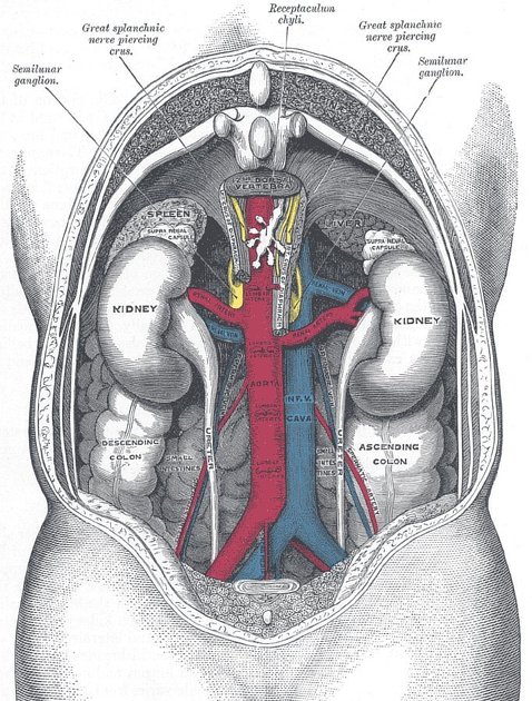
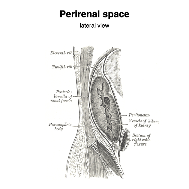
The perirenal space is the largest of the three divisions of the retroperitoneum and is the most easily identified. It contains the kidneys, renal vessels, proximal collecting systems, adrenal glands and an adequate amount of fat to allow identification on CT scanning. It also contains the perinephric bridging septa of Kunin 5.
The space is surrounded by the perirenal fascia and is in continuity with the opposite perirenal space across the midline. It abuts the bare area of the liver on the right and the subphrenic space on the left; there is mediastinal communication via the various diaphragmatic hiatus.
In disease, the space is usually closed inferiorly, preventing pelvic spread.
Related pathology
Lesions that may involve the perirenal space include 4:
References
- 1. Susan Standring. Gray's Anatomy. (2008) ISBN: 9780443066849 - Google Books
- 2. Michael Schünke, Lawrence M. Ross, Erik Schulte et al. General Anatomy and Musculoskeletal System. (2010) ISBN: 9781604062922 - Google Books
- 3. Anne M. Gilroy, Brian R. MacPherson, Lawrence M. Ross. Atlas of Anatomy. (2008) ISBN: 9781604060621 - Google Books
- 4. Westphalen A, Yeh B, Qayyum A, Hari A, Coakley F. Differential Diagnosis of Perinephric Masses on CT and MRI. AJR Am J Roentgenol. 2004;183(6):1697-702. doi:10.2214/ajr.183.6.01831697 - Pubmed
- 5. Kunin M. Bridging Septa of the Perinephric Space: Anatomic, Pathologic, and Diagnostic Considerations. Radiology. 1986;158(2):361-5. doi:10.1148/radiology.158.2.3941862 - Pubmed
Incoming Links
- Adrenal gland
- Urinoma
- Renal abscess
- Lateroconal fascia
- Abdominal aortic aneurysm rupture
- Wunderlich syndrome
- Perinephric bridging septa
- Extra-adrenal myelolipoma
- Retroperitoneum
- Perinephric abscess
- Renal fascia
- Great vessel space
- Perinephric stranding
- Renal sinus
- Erdheim-Chester disease
- Kidneys
- Perirenal cobweb
- Urogenital curriculum
- Perirenal space (Gray's illustrations)
- Wunderlich syndrome
- Retroperitoneal spaces
- Renal artery thrombosis
- Renal cell carcinoma
- Renal cell carcinoma
- Perinephric lymphoma
- Spontaneous subcapsular renal hematoma
- Perirenal/perihilar follicular lymphoma
- B cell lymphoma - perirenal
- Retroperitoneum
- Infected perinephric haematoma
- IVC filter induced perinephric haematoma
- Perinephric abscess pointing into the posterior chest wall
- Perirenal haematoma - post biopsy
- CT guided renal biopsy - post procedure perirenal haematoma
- Emphysematous pyelonephritis
- Urinoma
- Retroperitoneal abscess
Related articles: Anatomy: Abdominopelvic
- skeleton of the abdomen and pelvis
- muscles of the abdomen and pelvis
- spaces of the abdomen and pelvis
- anterior abdominal wall
- posterior abdominal wall
- abdominal cavity
- pelvic cavity
- perineum
- abdominal and pelvic viscera
- gastrointestinal tract
- spleen
- hepatobiliary system
-
endocrine system
-
adrenal gland
- adrenal vessels
- chromaffin cells
- variants
- pancreas
- organs of Zuckerkandl
-
adrenal gland
-
urinary system
-
kidney
- renal pelvis
- renal sinus
- avascular plane of Brodel
-
variants
- number
- fusion
- location
- shape
- ureter
- urinary bladder
- urethra
- embryology
-
kidney
- male reproductive system
-
female reproductive system
- vulva
- vagina
- uterus
- adnexa
- Fallopian tubes
- ovaries
- broad ligament (mnemonic)
- variant anatomy
- embryology
- blood supply of the abdomen and pelvis
- arteries
-
abdominal aorta
- inferior phrenic artery
- celiac artery
- superior mesenteric artery
- middle suprarenal artery
- renal artery (variant anatomy)
- gonadal artery (ovarian artery | testicular artery)
- inferior mesenteric artery
- lumbar arteries
- median sacral artery
-
common iliac artery
- external iliac artery
-
internal iliac artery (mnemonic)
- anterior division
- umbilical artery
- superior vesical artery
- obturator artery
- vaginal artery
- inferior vesical artery
- uterine artery
- middle rectal artery
-
internal pudendal artery
- inferior rectal artery
-
perineal artery
- posterior scrotal artery
- transverse perineal artery
- artery to the bulb
- deep artery of the penis/clitoris
- dorsal artery of the penis/clitoris
- inferior gluteal artery
- posterior division (mnemonic)
- variant anatomy
- anterior division
-
abdominal aorta
- portal venous system
- veins
- anastomoses
- arterioarterial anastomoses
- portal-systemic venous collateral pathways
- watershed areas
- arteries
- lymphatics
- innervation of the abdomen and pelvis
- thoracic splanchnic nerves
- lumbar plexus
-
sacral plexus
- lumbosacral trunk
- sciatic nerve
- superior gluteal nerve
- inferior gluteal nerve
- nerve to piriformis
- perforating cutaneous nerve
- posterior femoral cutaneous nerve
- parasympathetic pelvic splanchnic nerves
- pudendal nerve
- nerve to quadratus femoris and inferior gemellus muscles
- nerve to internal obturator and superior gemellus muscles
- autonomic ganglia and plexuses


 Unable to process the form. Check for errors and try again.