
Tarsal coalition describes the complete or partial union between two or more bones in the midfoot and hindfoot. Tarsal coalition refers to developmental fusion rather than fusion that is acquired secondary to conditions such as rheumatoid arthritis, trauma or post-surgical.
On this page:
Epidemiology
It occurs in ~5% of the population and although congenital, patients typically present in adolescence. There is a significant male predilection (M:F 4:1) 5. 50% are bilateral (even if symptomatic only on one side). Pes planus (flat foot) is usually a feature.
Clinical presentation
Many patients with tarsal coalition are asymptomatic. Clinical presentation is usually delayed until adolescence and is associated with either increased mechanical strain on the joints/structures on either side of the coalition or with progressive ossification of the coalition 3,5. The average age of clinical symptoms onset is lower in the case of calcaneonavicular coalition (8-12 years) than of talocalcaneal (12-16 years) because of earlier ossification of the former 3.
The condition is bilateral in up to 50% of cases, and clinical presentation includes:
- hindfoot or tarsal pain/stiffness
- tarsal tunnel syndrome
- peroneal tendon spasm
- pes planus
- formation of a ball and socket tibiotalar joint
- secondary osteoarthritis
Pathology
Tarsal coalition is believed to be the result of incomplete or faulty segmentation during development. They may be of three types, depending on the tissue which bridges between the two bones. The three types are 1:
- bony: synostosis
- cartilaginous: synchondrosis
- fibrous: syndesmosis
Associations
Radiographic features
The vast majority (90%) of tarsal coalitions are either:
-
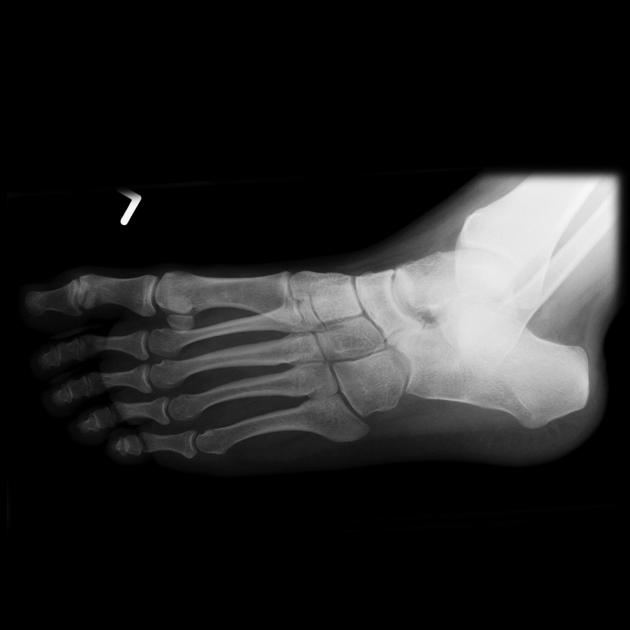
calcaneonavicular (~45%)
- usually involves the anterior process of the calcaneus
- maybe associated with hypoplasia of the talus
- best seen on oblique radiographs
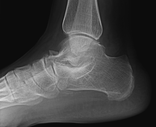
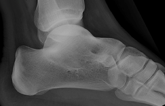
- the anteater nose sign may sometimes be seen on a lateral radiograph
-
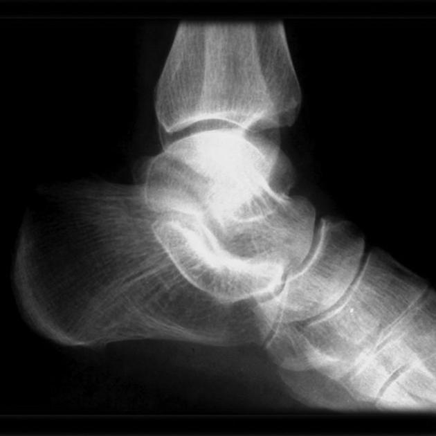
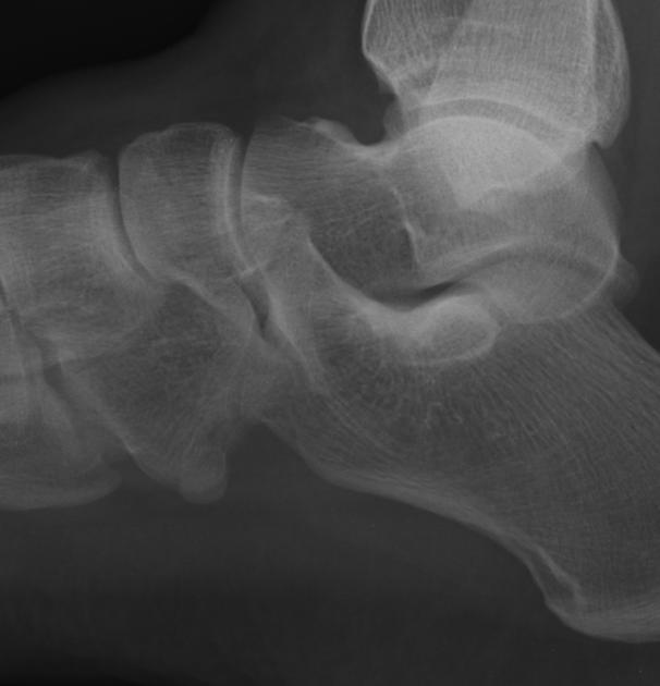
talocalcaneal (~45%)
- usually involves the middle facet
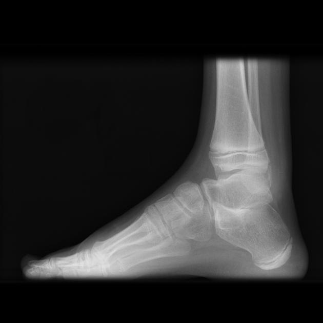
- best seen on the lateral view
- C-sign: complete posterior ring around the talus and sustentaculum tali
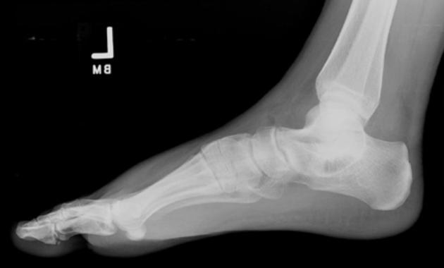
- talar beak sign due to impaired subtalar movement
The remainder of the coalitions (calcaneocuboid, talonavicular, cubonavicular) are much less common 3. The metatarsal-cuneiform coalition is a rare cause of midfoot pain.
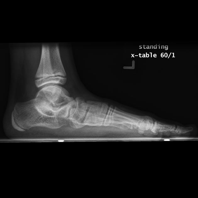
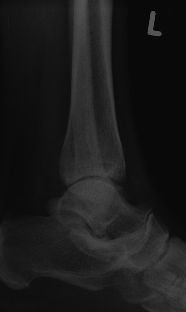
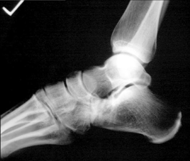
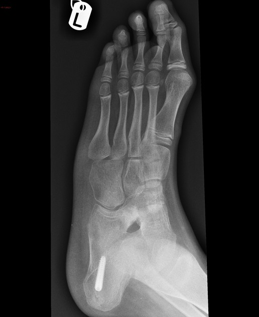
Plain radiograph
Plain films (AP + lateral + 45° internal oblique) are usually the first investigation of choice, and in many instances yield that diagnosis, especially in the case of calcaneonavicular and talonavicular coalition. Plain film findings are most apparent when there is a bony coalition, especially where the bony bar is large enough and the medullary cavity is seen in continuity between the two bones. In fibrous coalitions, there is irregularity and narrowing of the bony interfaces, and there is usually associated sclerosis.
General features of the non-osseous coalition include:
- subchondral reactive bony changes
- adjacent marrow edema (MR)
- unusual articular orientation
- joint space loss
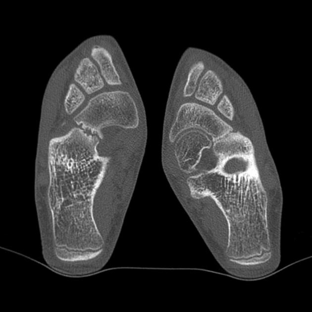
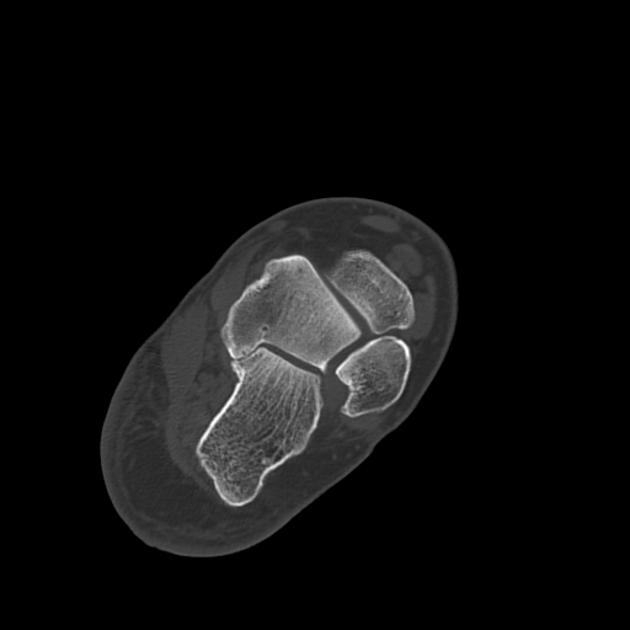
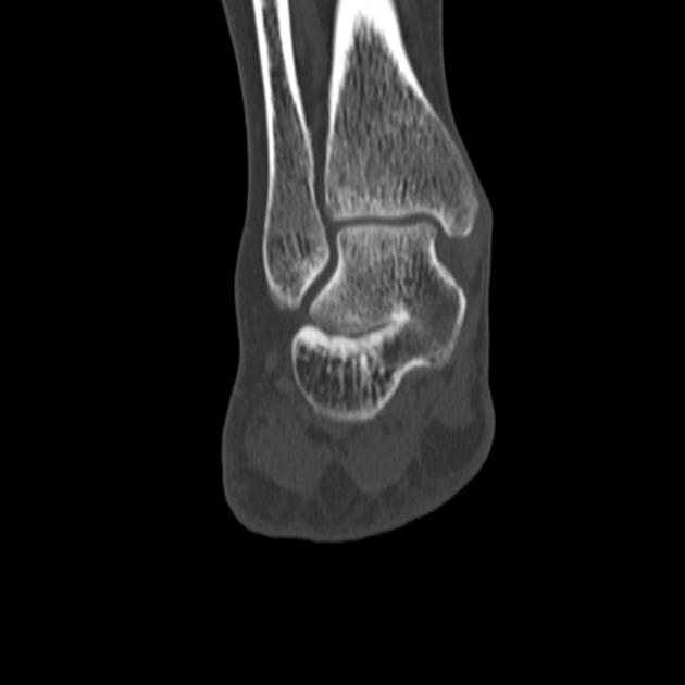
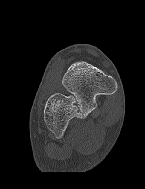
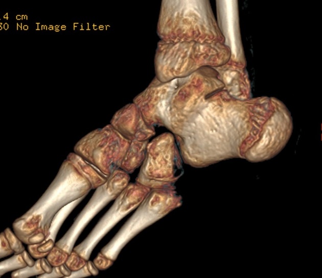
CT
Cross-sectional imaging (CT or MRI) is often needed in the case of talocalcaneal coalition due to the lower sensitivity of conventional radiography in its detection.
CT depicts precisely the extent of the coalition and is therefore useful in resection planning or determining the indications for arthrodesis.
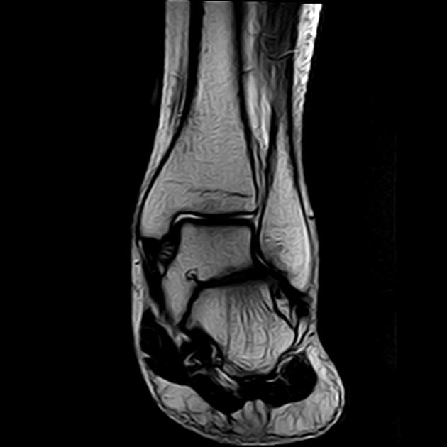
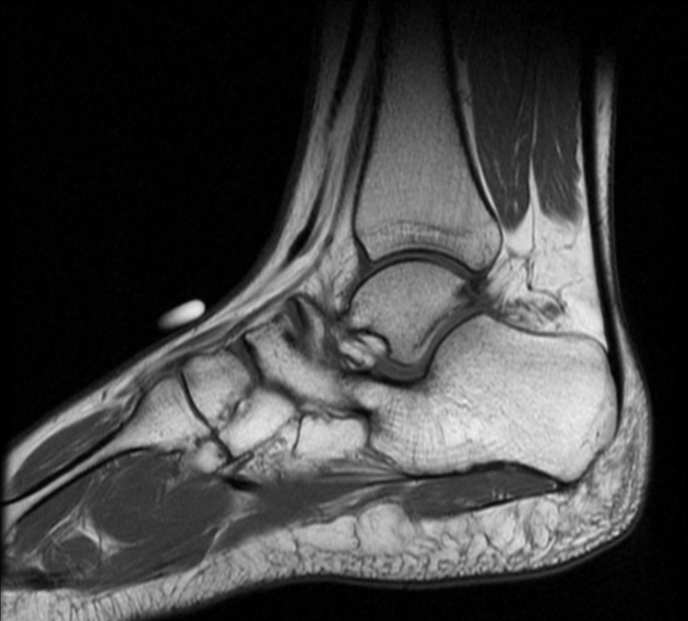
MRI
May help to distinguish between:
- osseous coalition
- continuity of marrow signal
- fibrous coalition
- proximity of surfaces with loss of the fat plane
- low signal on T1/T2 weighted sequences
- cartilaginous coalition
- proximity of surfaces with loss of the fat plane
- intermediate T2/STIR +/- fluid signal
Possible associated findings include marrow or soft tissue edema
Treatment and prognosis
Although conservative (non-surgical) therapy may be useful in alleviating pain, symptomatic improvement is usually short-lived since it does not address the underlying anatomic abnormality 6.
Surgical management usually involves an osteotomy and removal of the whole coalition. Space is then filled with soft tissues (e.g. muscle or fat). In case of resection failure or coexisting severe degenerative joint disease triple arthrodesis is usually performed, or alternatively (in case of a subtalar coalition) subtalar fusion 3.
History and etymology
A tarsal coalition has been first reported by Georges-Louis Leclerc, Comte de Buffon in 1769 8.


 Unable to process the form. Check for errors and try again.