
Tibialis anterior muscle
Citation, DOI, disclosures and article data
At the time the article was created Jeremy Jones had no recorded disclosures.
View Jeremy Jones's current disclosuresAt the time the article was last revised Arlene Campos had no financial relationships to ineligible companies to disclose.
View Arlene Campos's current disclosures- Tibialis anterior
- Tibialis anterior tendon
- Tibialis anterior (TAT)
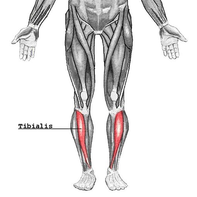
The tibialis anterior muscle is a long narrow fusiform-shaped muscle located in the anterior compartment of the leg. It is the most superficial and largest muscle of the group and is the main foot dorsiflexor.
Summary
origin: superior two thirds of the lateral surface of tibia and adjacent interosseous membrane
insertion: inferomedial aspect of medial cuneiform and base of first metatarsal
-
action:
dorsiflexion of foot at ankle
inversion of the foot (at subtalar and midtarsal joints)
helps to hold up medial longitudinal arch of foot
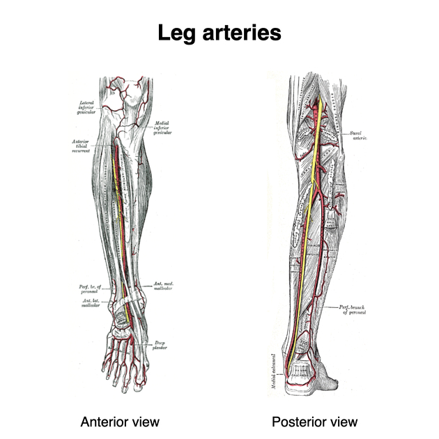
blood supply: branches of the anterior tibial and fibular arteries
innervation: deep peroneal nerve (L4 - S1)
On this page:
Gross anatomy
Relations
The tibialis anterior lies medial to the extensor digitorum longus and extensor hallucis longus. The muscle pierces the superior extensor retinacula but travels deep to the inferior retinacula which holds it in place.
Origin
The tibialis anterior muscle originates from the lateral condyle and upper two-thirds of the lateral surface of the tibia. Additionally, it arises from the interosseous membrane of the leg, deep surface of the fascia cruris and the anterior intermuscular septum.
Insertion
The tibialis anterior inserts on the medial cuneiform bone and base of the first metatarsal bone.
Action
The tibialis anterior muscle provides dorsiflexion at the talocrural (ankle) joint and inversion of the foot at the subtalar and midtarsal (Chopart's) joints. It also plays a role in supporting the medial longitudinal arch of the foot.
Innervation
The tibialis anterior is innervated by the deep peroneal (fibular) nerve (L4 - S1), a branch of the common peroneal nerve.
Arterial supply
The muscle receives arterial blood supply primarily from the branches of the anterior tibial artery in addition to branches of the posterior tibial artery.
Variant anatomy
Os sesamoideum tibialis anterioris is an accessory sesamoid bone that arises during embryonic development which is sometimes incorrectly misdiagnosed as a fracture 4.
References
- 1. Susan Standring. Gray's Anatomy. (2008) ISBN: 9780443066849 - Google Books
- 2. Varghese A, Bianchi S. Ultrasound of tibialis anterior muscle and tendon: anatomy, technique of examination, normal and pathologic appearance. J Ultrasound. 2014;17 (2): 113-23. doi:10.1007/s40477-013-0060-7 - Free text at pubmed - Pubmed citation
- 3. Mengiardi B, Pfirrmann C, Vienne P et al. Anterior Tibial Tendon Abnormalities: MR Imaging Findings. Radiology. 2005;235(3):977-84. doi:10.1148/radiol.2353040617 - Pubmed
- 4. Theodore Eliot Keats, Mark Anderson. Atlas of Normal Roentgen Variants That May Simulate Disease. (2001) ISBN: 9780323013222 - Google Books
Incoming Links
- Anterior compartment of the leg
- Sciatic nerve motor distribution
- Extensor digitorum longus muscle
- Tibioastragalus anticus of Gruber muscle
- Deep peroneal nerve entrapment
- Fibularis longus muscle
- Cuneonavicular joint
- Anterior medial malleolar artery
- Plantaris muscle
- Ankle extensor compartment injury
- Tibialis posterior muscle
- Gastrocnemius muscle
- Muscles of the lower limb
- Metatarsal
- Extensor hallucis longus muscle
- Extensor hallucis capsularis tendon
- Tibia
- Inclusion body myositis
- Ankle joint
- Gerdy tubercle
- Insertional tendinopathy of the tibialis anterior tendon
- Muscle hernias - tibialis anterior muscle
- Foreign body - leg
- Tibialis anterior tendon laceration
- Tibialis anterior tendon bony avulsion
- Extensor digitorum longus tenosynovitis
- Muscle hernia - leg
- Tibialis anterior muscle hernia
- Tibialis anterior tendon rupture - atraumatic
- Foreign body (ankle)
- Foreign body - distal leg
- Foot muscle insertion - anterior (Gray's illustration)
- Tibialis anterior muscle (illustration)
- Foot muscle insertions (Gray's anatomy illustration)
- Tibialis anterior tenosynovitis
- Tibialis anterior tendinosis
- Tibialis anterior tendon rupture
- MRI anatomy of ankle
Related articles: Anatomy: Lower limb
- skeleton of the lower limb
- joints of the lower limb
-
hip joint
- ligaments
- muscles
- additional structures
- hip joint capsule
- zona orbicularis
- iliotibial band
-
hip bursae
- anterior
- iliopsoas bursa (iliopectineal bursa)
- lateral
- subgluteal bursae
- greater trochanteric bursa (subgluteus maximus bursa)
- subgluteus medius bursa
- subgluteus minimus bursa
- gluteofemoral bursa
- subgluteal bursae
- postero-inferior
- anterior
- ossification centers
-
knee joint
- ligaments
- anterior cruciate ligament
- posterior cruciate ligament
- medial collateral ligament
- lateral collateral ligament
- meniscofemoral ligament (mnemonic)
-
posterolateral ligamentous complex
- arcuate ligament
- patellar tendon and quadriceps tendon
- anterolateral ligament
- posterior oblique ligament
- oblique popliteal ligament
- medial patellofemoral ligament
- additional structures
- extensor mechanism of the knee
- groove for the popliteus tendon
- knee bursae
- anterior bursae
- medial bursae
- lateral bursae
- posterior bursae
- knee capsule
- lateral patellar retinaculum
- medial patellar retinaculum
- menisci
- pes anserinus (mnemonic)
- ossification centers
- ligaments
- tibiofibular joints
-
ankle joint
- regional anatomy
- medial ankle
- lateral ankle
- anterior ankle
- ligaments
- medial collateral (deltoid) ligament
- lateral collateral ligament
- additional structures
- ankle bursae
- ossification centers of the ankle
- variants
- regional anatomy
- foot joints
- subtalar joint
- mid-tarsal (Chopart) joint
-
tarsometatarsal (Lisfranc) joint
- ligaments
- intermetatarsal joint
- metatarsophalangeal joint
- interphalangeal joint
- ossification centers
-
hip joint
- spaces of the lower limb
-
muscles of the lower limb
- muscles of the pelvic group
- muscles of the thigh
- muscles of the leg
- anterior compartment of the leg
- posterior compartments of the leg
- lateral compartment of the leg
- muscles of the foot
- dorsal muscles
- plantar muscles
- 1st layer
- 2nd layer
- 3rd layer
- 4th layer
- accessory muscles of the lower limb
- accessory gluteal muscles
-
accessory muscles of the ankle
- accessory peroneal muscles
- accessory flexor digitorum longus muscle
- accessory soleus muscle
- peroneocalcaneus internus muscle
- tibiocalcaneus internus muscle
- extensor hallucis capsularis tendon
- anterior fibulocalcaneus muscle
- accessory extensor digiti secundus muscle
- tibioastragalus anticus of Gruber muscle
- vascular supply of the lower limb
- arterial supply of the lower limb
- venous drainage of the lower limb
- innervation of the lower limb
- lymphatic system of the lower limb
- lymphatic pathways
- anteromedial group
- anterolateral group
- posteromedial group
- posterolateral group
- lower limb lymph nodes
- lymphatic pathways


 Unable to process the form. Check for errors and try again.