
Umbilical artery
Citation, DOI, disclosures and article data
At the time the article was created Grace Florescu had no recorded disclosures.
View Grace Florescu's current disclosuresAt the time the article was last revised Craig Hacking had the following disclosures:
- Philips Australia, Paid speaker at Philips Spectral CT events (ongoing)
These were assessed during peer review and were determined to not be relevant to the changes that were made.
View Craig Hacking's current disclosures- Umbilical arteries
The umbilical artery gives rise to both a nonfunctional remnant of the fetal circulation and an active vessel giving supply to the bladder. In the adult, the obliterated area of the vessel is identifiable as the medial umbilical ligament and the patent segment is the superior vesical artery.
Summary
- origin: anterior division of the internal iliac artery
- location: abdominal wall and pelvis
- supply: placenta in the fetus, superior bladder, ureter, ductus deferens
- main branches: obliterated umbilical artery, superior vesical artery
Gross anatomy
Origin
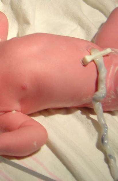
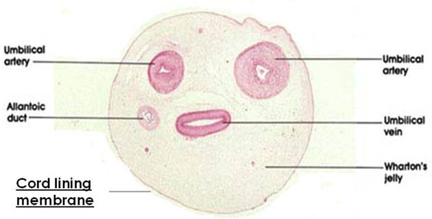
The umbilical artery originates from the anterior division of the internal iliac artery. In the fetus, it travels within the umbilical cord to the placenta and hence is the communication to the maternal circulation.
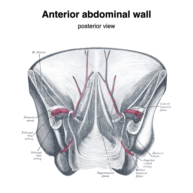
After the umbilical cord has been cut at birth, clots form within the vessel and it obliterates. Its remnant can be found in the posterior aspect of the anterior abdominal wall as the medial umbilical ligament, which courses superomedially to the umbilicus, within a fold of peritoneum called the medial umbilical fold. It is a paired structure and lies lateral to the median umbilical ligament (the urachal remnant) and medial to the lateral umbilical fold, which contains the inferior epigastric vessels.
Branches
Within the fetus, the artery terminates in the placenta as multiple chorionic vessels.
In the adult, there are two vessels which the umbilical artery gives rise to; these are not branches but are actually a direct continuation of the umbilical artery:
- obliterated umbilical artery: this is the distal part of the umbilical artery that thromboses soon after birth and hence becomes obliterated; it may be found as a remnant seen in the internal aspect of the anterior abdominal wall
- superior vesical artery: the persistent proximal portion of the umbilical artery; runs along the side wall of the pelvis to its supply at the superior bladder as well as adjacent ureter and ductus deferens
Supply
In the fetus, the paired umbilical arteries travel within the umbilical cord to carry deoxygenated blood from the fetus to the mother.
In the adult, the superior vesical artery gives vascular supply to the superior bladder, the portion of the ureter next to this and the ductus deferens.
Quiz questions
References
- 1. Last's anatomy, regional and applied. Churchill Livingstone. ISBN:044304662X. Read it at Google Books - Find it at Amazon
- 2, Dalley AF, Agur AM. Clinically Oriented Anatomy, Sixth Edition. Lippincott Williams & Wilkins. ISBN:1605476528. Read it at Google Books - Find it at Amazon
- 3. Gray's Anatomy. Churchill Livingstone. (2011) ISBN:0443066841. Read it at Google Books - Find it at Amazon
Incoming Links
- Branches of internal iliac artery (mnemonic)
- Fetal circulation
- Umbilical cord
- Umbilical cord thrombosis
- Superior vesical artery
- Placenta
- Umbilical arterial Doppler assessment
- Single umbilical artery
- Medial umbilical folds
- Branches of anterior and posterior divisions of the internal iliac artery (mnemonic)
- Second trimester ultrasound scan
- Internal iliac artery
- Medical abbreviations and acronyms (U)
- Branches of the anterior division of the internal iliac artery (mnemonic)
- Umbilical arterial catheters
- Umbilicus
- Umbilical vein
- Fetal urinary bladder
- Inferior hypogastric plexus
- Absent umbilical arterial end-diastolic flow
Related articles: Anatomy: Abdominopelvic
- skeleton of the abdomen and pelvis
- muscles of the abdomen and pelvis
- spaces of the abdomen and pelvis
- anterior abdominal wall
- posterior abdominal wall
- abdominal cavity
- pelvic cavity
- perineum
- abdominal and pelvic viscera
- gastrointestinal tract
- spleen
- hepatobiliary system
-
endocrine system
-
adrenal gland
- adrenal vessels
- chromaffin cells
- variants
- pancreas
- organs of Zuckerkandl
-
adrenal gland
-
urinary system
-
kidney
- renal pelvis
- renal sinus
- avascular plane of Brodel
-
variants
- number
- fusion
- location
- shape
- ureter
- urinary bladder
- urethra
- embryology
-
kidney
- male reproductive system
-
female reproductive system
- vulva
- vagina
- uterus
- adnexa
- Fallopian tubes
- ovaries
- broad ligament (mnemonic)
- variant anatomy
- embryology
- blood supply of the abdomen and pelvis
- arteries
-
abdominal aorta
- inferior phrenic artery
- celiac artery
- superior mesenteric artery
- middle suprarenal artery
- renal artery (variant anatomy)
- gonadal artery (ovarian artery | testicular artery)
- inferior mesenteric artery
- lumbar arteries
- median sacral artery
-
common iliac artery
- external iliac artery
-
internal iliac artery (mnemonic)
- anterior division
- umbilical artery
- superior vesical artery
- obturator artery
- vaginal artery
- inferior vesical artery
- uterine artery
- middle rectal artery
-
internal pudendal artery
- inferior rectal artery
-
perineal artery
- posterior scrotal artery
- transverse perineal artery
- artery to the bulb
- deep artery of the penis/clitoris
- dorsal artery of the penis/clitoris
- inferior gluteal artery
- posterior division (mnemonic)
- variant anatomy
- anterior division
-
abdominal aorta
- portal venous system
- veins
- anastomoses
- arterioarterial anastomoses
- portal-systemic venous collateral pathways
- watershed areas
- arteries
- lymphatics
- innervation of the abdomen and pelvis
- thoracic splanchnic nerves
- lumbar plexus
-
sacral plexus
- lumbosacral trunk
- sciatic nerve
- superior gluteal nerve
- inferior gluteal nerve
- nerve to piriformis
- perforating cutaneous nerve
- posterior femoral cutaneous nerve
- parasympathetic pelvic splanchnic nerves
- pudendal nerve
- nerve to quadratus femoris and inferior gemellus muscles
- nerve to internal obturator and superior gemellus muscles
- autonomic ganglia and plexuses


 Unable to process the form. Check for errors and try again.