
Volar plate avulsion injury
Citation, DOI, disclosures and article data
At the time the article was created Charlie Chia-Tsong Hsu had no recorded disclosures.
View Charlie Chia-Tsong Hsu's current disclosuresAt the time the article was last revised Mohammad Taghi Niknejad had no financial relationships to ineligible companies to disclose.
View Mohammad Taghi Niknejad's current disclosures- Volar plate avulsion injuries
- Volar plate avulsion fracture
- Volar plate avulsion fractures
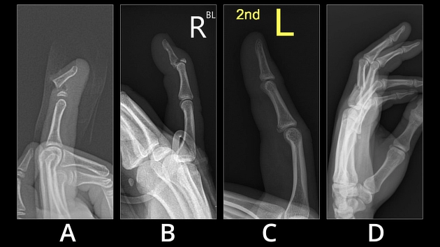
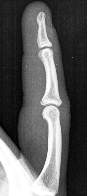
Volar plate avulsion injuries are a type of avulsion injury. The volar plate of the proximal interphalangeal (PIP) joint is vulnerable to hyperextension injury, in the form of either a ligament tear or an intra-articular fracture.
On this page:
Gross anatomy
The volar plate forms the floor of the PIP joint separating the joint space from the flexor tendon sheath. The volar plate has a ligamentous origin on the proximal phalanx with a capsular insertion onto the middle phalanx.
Pathology
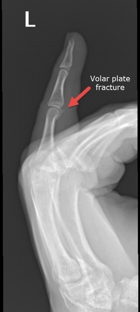
Hyperextension injury involving the PIP of the finger can avulse the volar plate which is commonly associated with a volar avulsion fracture at the base of the middle phalanx.
When the volar avulsion fracture involves a significant portion of the articular surface, instability and dorsal dislocation of middle phalanx can occur. This is because a greater portion of the stabilizing collateral ligaments is attached to the avulsed fragment.
Classification
Two classification systems are considered most useful for the management of these injuries 7:
Radiographic features
Plain radiography / CT
A small fragment of bone is avulsed from the volar base of the middle phalanx. If there is significant involvement of the articular surface, this may be associated with dorsal dislocation of the middle phalanx.
Ultrasound
May be seen as a small displaced echogenic focus. High-resolution sonography may be useful evaluate palmar plate stability and to assess reduction of edema especially in an acute setting 6.
Treatment and prognosis
Overall, a small fracture fragment (<40% articular surface) and/or reducible fracture with <30 degrees of flexion is usually managed conservatively with finger splinting 7. A large fragment (>40% articular surface) or >30 degrees of flexion to reduce the fragment and malalignment post-closed reduction are indicators for operative treatment 7.
References
- 1. Nance E, Kaye J, Milek M. Volar Plate Fractures. Radiology. 1979;133(1):61-4. doi:10.1148/133.1.61 - Pubmed
- 2. Borut Marincek, Robert F. Dondelinger. Emergency Radiology. (2006) ISBN: 9783540262275 - Google Books
- 3. Eaton R & Malerich M. Volar Plate Arthroplasty of the Proximal Interphalangeal Joint: A Review of Ten Years' Experience. J Hand Surg Am. 1980;5(3):260-8. doi:10.1016/s0363-5023(80)80011-6 - Pubmed
- 4. Pattni A, Jones M, Gujral S. Volar Plate Avulsion Injury. Eplasty. 2016;16:ic22. PMC4894366 - Pubmed
- 5. Wieschhoff G, Sheehan S, Wortman J et al. Traumatic Finger Injuries: What the Orthopedic Surgeon Wants to Know. Radiographics. 2016;36(4):1106-28. doi:10.1148/rg.2016150216 - Pubmed
- 6. Leclère F, Mathys L, Juon B, Vögelin E. The Role of Dynamic Ultrasound in the Immediate Conservative Treatment of Volar Plate Injuries of the PIP Joint: A Series of 78 Patients. Plast Surg (Oakv). 2017;25(3):151-6. doi:10.1177/2292550317716122 - Pubmed
- 7. Pattni A, Jones M, Gujral S. Volar Plate Avulsion Injury. Eplasty. 2016;16:ic22. PMC4894366 - Pubmed
Incoming Links
- Volar plate avulsion fracture
- Volar plate avulsion injury
- Proximal interphalangeal joint dislocation - 3rd and 4th fingers
- Proximal interphalangeal joint dislocation - 3rd finger
- PIP joint dislocation with volar plate avulsion fracture
- Fracture of the ulnar sesamoid of the first metacarpophalangeal joint
- Volar plate avulsion injury
- Volar plate avulsion fracture
- Volar plate avulsion injury
- Volar plate avulsion injury
- Volar plate avulsion injury
- Volar plate avulsion injury - subtle
- Proximal interphalangeal joint (PIP) dislocation with avulsion tear of the volar plate
- Fracture base of proximal phalanx of index finger
- Volar plate avulsion fracture
- Volar plate avulsion injury
- Volar plate avulsion injury
Related articles: Fractures
-
fracture
- terminology[+][+]
- fracture location
- diaphyseal fracture
- metaphyseal fracture
- physeal fracture
- epiphyseal fracture
- fracture types
- avulsion fracture
- articular surface injuries
- complete fracture
- incomplete fracture
- infraction
- compound fracture
- pathological fracture
- stress fracture
- fracture displacement
- fracture location
- fracture healing[+][+]
- skull fractures[+][+]
-
facial fractures[+][+]
- fractures involving a single facial buttress
- alveolar process fractures
- frontal sinus fracture
- isolated zygomatic arch fractures
- mandibular fracture
- nasal bone fracture
- orbital blow-out fracture
- paranasal sinus fractures
- complex fractures
- dental fractures
- fractures involving a single facial buttress
-
spinal fractures[+][+]
- classification (AO Spine classification systems)
-
cervical spine fracture classification systems
- AO classification of upper cervical injuries
- AO classification of subaxial injuries
- Anderson and D'Alonzo classification (odontoid fracture)
- Roy-Camille classification (odontoid process fracture)
- Gehweiler classifcation (atlas fractures)
- Levine and Edwards classification (hangman fracture)
- Allen and Ferguson classification (subaxial spine injuries)
- subaxial cervical spine injury classification (SLIC)
- thoracolumbar spinal fracture classification systems
- three column concept of spinal fractures (Denis classification)
- classification of sacral fractures
-
cervical spine fracture classification systems
- spinal fractures by region
- spinal fracture types
- classification (AO Spine classification systems)
- rib fractures[+][+]
- sternal fractures
-
upper limb fractures
- classification[+][+]
- Rockwood classification (acromioclavicular joint injury)
- AO classification (clavicle fracture)
- Neer classification (clavicle fracture)
- Neer classification (proximal humeral fracture)
- AO classification (proximal humeral fracture)
- AO/OTA classification of distal humeral fractures
- Milch classification (lateral humeral condyle fracture)
- Weiss classification (lateral humeral condyle fracture)
- Bado classification of Monteggia fracture-dislocations (radius-ulna)
- Mason classification (radial head fracture)
- Frykman classification (distal radial fracture)
- Mayo classification (scaphoid fracture)
- Hintermann classification (gamekeeper's thumb)
- Eaton classification (volar plate avulsion injury)
- Keifhaber-Stern classification (volar plate avulsion injury)
- upper limb fractures by region
- shoulder[+][+]
- clavicular fracture
-
scapular fracture
- acromion fracture
- coracoid process fracture
- glenoid fracture
- humeral head fracture
- proximal humeral fracture
- humeral neck fracture
- arm[+][+]
- elbow[+][+]
- forearm[+][+]
- wrist[+][+]
-
carpal bones[+][+]
- scaphoid fracture
- lunate fracture
- capitate fracture
- triquetral fracture
- pisiform fracture
- hamate fracture
- trapezoid fracture
- trapezium fracture
- hand
- shoulder[+][+]
- classification[+][+]
- lower limb fractures[+][+]
- classification by region
- pelvic fractures
- hip fractures
- Pipkin classification (femoral head fracture)
- Garden classification (hip fracture)
- American Academy of Orthopedic Surgeons classification (periprosthetic hip fracture)
- Cooke and Newman classification (periprosthetic hip fracture)
- Johansson classification (periprosthetic hip fracture)
- Vancouver classification (periprosthetic hip fracture)
- femoral
- knee
- Schatzker classification (tibial plateau fracture)
- AO classification of distal femur fractures
- Meyers and McKeevers classification (anterior cruciate ligament avulsion fracture)
- tibia/fibula
- Watson-Jones classification (tibial tuberosity avulsion fracture)
- ankle
- foot
- Berndt and Harty classification (osteochondral lesions of the talus)
- Sanders CT classification (calcaneal fracture)
- Hawkins classification (talar neck fracture)
- Myerson classification (Lisfranc injury)
- Nunley-Vertullo classification (Lisfranc injury)
- pelvis and lower limb fractures by region
- pelvic fracture
- sacral fracture
- coccygeal fracture
-
hip
- acetabular fracture
- femoral head fracture
-
femoral neck fracture
- subcapital fracture
- transcervical fracture
- basicervical fracture
-
trochanteric fracture
- pertrochanteric fracture
- intertrochanteric fracture
- subtrochanteric fracture
- femur
- mid-shaft fracture
- bisphosphonate-related fracture
- distal femoral fracture
- knee
- avulsion fractures
- Segond fracture
- reverse Segond fracture
- anterior cruciate ligament avulsion fracture
- posterior cruciate ligament avulsion fracture
- arcuate complex avulsion fracture (arcuate sign)
- biceps femoris avulsion fracture
- iliotibial band avulsion fracture
- semimembranosus tendon avulsion fracture
- Stieda fracture (MCL avulsion fracture)
- patellar fracture
- tibial plateau fracture
- avulsion fractures
- leg
- tibial tuberosity avulsion fracture
- tibial shaft fracture
- fibular shaft fracture
- Maisonneuve fracture
- ankle
- foot
- tarsal bones
- metatarsal bones
- phalanges
- classification by region
- terminology[+][+]


 Unable to process the form. Check for errors and try again.