Tibial plateau fracture
Citation, DOI, disclosures and article data
At the time the article was created The Radswiki had no recorded disclosures.
View The Radswiki's current disclosuresAt the time the article was last revised Tariq Walizai had no financial relationships to ineligible companies to disclose.
View Tariq Walizai's current disclosures- Tibial plateau fractures
- Fractures of the tibial plateau
Tibial plateau fractures were originally termed a bumper fracture or fender fracture but only 25% of tibial plateau fractures result from impact with automobile bumpers.
On this page:
Pathology
The most common mechanism of injury involves axial loading, e.g. fall from a significant height. In younger patients, the most common pattern of fracture is splitting, while in older, more osteoporotic patients, depression fractures typically are sustained.
Soft tissue injuries (e.g. to cruciate and collateral ligaments) occur in approximately 10% of patients.
Fractures of the lateral plateau are much more common than the medial plateau. To injure the medial plateau, a large amount of force is required; fractures of the medial plateau are usually seen in conjunction with fractures of the lateral plateau and other bones around the knee joint.
The fracture pattern will depend on the mechanism of injury. The Schatzker classification is a useful classification to categorize the mechanism of injury 1:
-
Schatzker I: wedge-shaped lateral plateau fracture
valgus force with axial loading (femoral condyle rams the tibial plateau)
-
Schatzker II: wedge-shaped lateral plateau fracture with compression fracture of ipsilateral plateau
valgus force (moderate association with medial collateral ligament and medial meniscus injury)
-
Schatzker III: compression fracture of the lateral plateau
axial force
-
Schatzker IV: medial plateau fracture with a split or compressed portion
varus force with axial load
-
Schatzker V: wedge fracture of both plateaus
complex high energy mechanism involving varus OR valgus forces with significant axial loading
-
Schatzker VI: transverse tibial metadiaphyseal fracture, along with any type of tibial plateau fracture
complex high energy mechanism involving varus OR valgus forces with significant axial loading
ADVERTISEMENT: Supporters see fewer/no ads
Radiographic features
Tibial plateau fractures are complex injuries that require adequate imaging to assess prior to fixation.
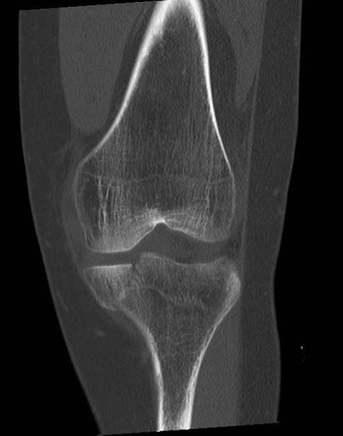
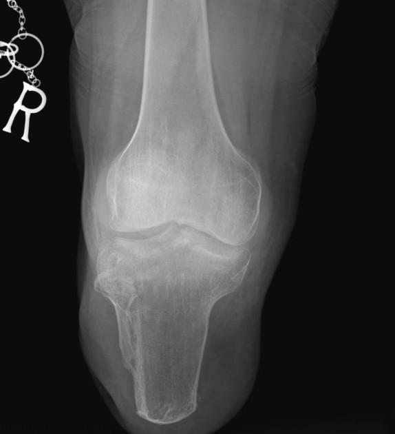
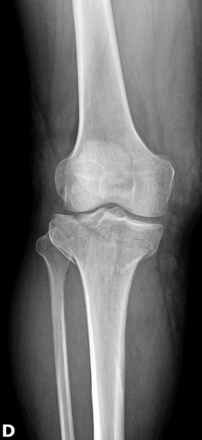
Plain radiograph
Plain radiography often underestimates the severity of the injury. Lipohemarthrosis should be present.
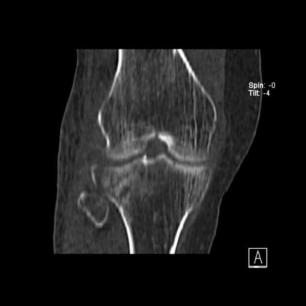
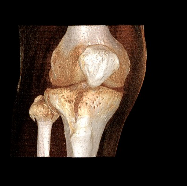
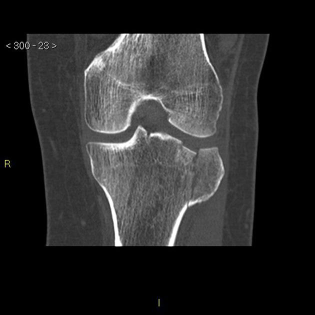
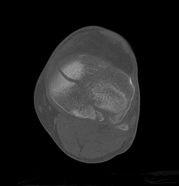
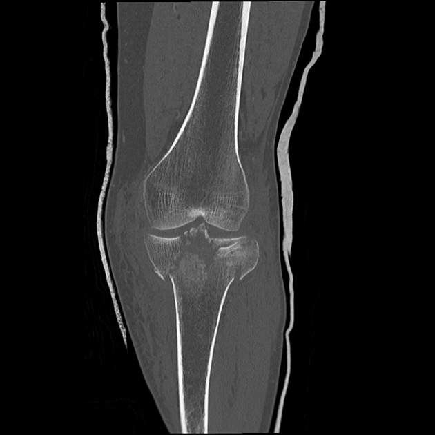
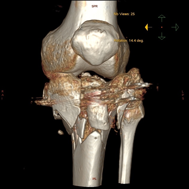
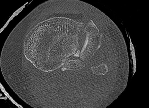
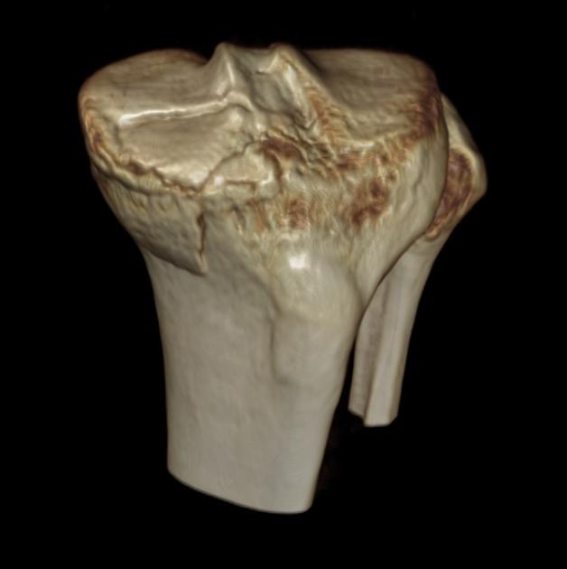
CT
CT is very helpful in accurately defining the extent of the bony injury and facilitates orthopedic intervention.
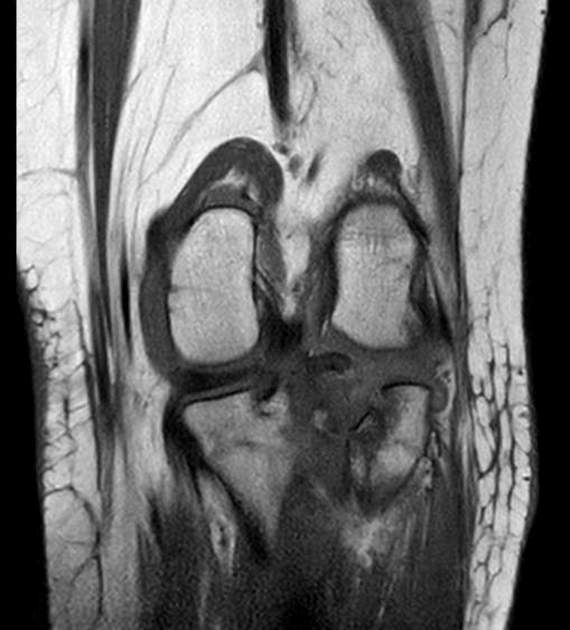
MRI
MRI is very helpful in the assessment of soft tissue injury around the joint.
Treatment and prognosis
The goal of therapy is to reduce the fracture and begin early mobilization. If the patient is immobilized for a lengthy period (>3 weeks), the joint will not return to the full range of movement.
Depression of a tibial plateau that is inadequately corrected results in a varus or valgus deformity and accelerated osteoarthritis.
Unappreciated ligamentous injury causes greater than normal stress on the remaining support structures of the joint, malalignment, and the development of premature osteoarthritis.
Quiz questions
References
- 1. Markhardt B, Gross J, Monu J. Schatzker Classification of Tibial Plateau Fractures: Use of CT and MR Imaging Improves Assessment1. Radiographics. 2009;29 (2): 585-597. Radiographics (full text) - doi:10.1148/rg.292085078
- 2. Macarini L, Murrone M, Marini S et-al. Tibial plateau fractures: evaluation with multidetector-CT. Radiol Med. 2005;108 (5-6): 503-14. Pubmed citation
- 3. Rafii M, Firooznia H, Golimbu C et-al. Computed tomography of tibial plateau fractures. AJR Am J Roentgenol. 1984;142 (6): 1181-6. AJR Am J Roentgenol (abstract) - Pubmed citation
- 4. Rafii M, Firooznia H, Golimbu C et-al. Computed tomography of tibial plateau fractures. AJR Am J Roentgenol. 1984;142 (6): 1181-6. AJR Am J Roentgenol (abstract) - Pubmed citation
Incoming Links
- Tibial plateau
- Paediatric knee radiograph (an approach)
- Tibial shaft fracture
- Knee joint
- Tibiofemoral joint
- Meniscal tear
- Schatzker classification of tibial plateau fractures
- Knee radiograph (an approach)
- Lower extremity fractures
- Lipohaemarthrosis
- Proximal tibiofibular joint dislocation
- Kfuri and Schatzker classification of tibial plateau fractures
- Tibial spine fracture
- Tibial plateau fracture - Schatzker type V
- Tibial plateau fracture
- Entrapped lateral meniscus in tibial plateau fracture
- Schatzker type V tibial plateau fracture with head of fibula fracture
- Tibial plateau fracture
- Kfuri and Schatzker classification of tibial plateau fractures
- Tibial plateau ORIF - Schatzker type III fracture
- Tibial plateau fracture - Schatzker type VI
- Tibial plateau fracture - Schatzker type II
- Tibial plateau fracture - Schatzker type IV
- Tibial plateau fracture - Schatzker type V
- Tibial plateau fracture - Schatzker type VI
- Schatzker classification of tibial plateau fractures (illustrations)
- Segond fracture
- Lateral tibial plateau fracture with lipohaemarthrosis
- Tibial plateau fracture
- Schatzker type II fracture
Related articles: Fractures
-
fracture
- terminology[+][+]
- fracture location
- diaphyseal fracture
- metaphyseal fracture
- physeal fracture
- epiphyseal fracture
- fracture types
- avulsion fracture
- articular surface injuries
- complete fracture
- incomplete fracture
- infraction
- compound fracture
- pathological fracture
- stress fracture
- fracture displacement
- fracture location
- fracture healing[+][+]
- skull fractures[+][+]
-
facial fractures[+][+]
- fractures involving a single facial buttress
- alveolar process fractures
- frontal sinus fracture
- isolated zygomatic arch fractures
- mandibular fracture
- nasal bone fracture
- orbital blow-out fracture
- paranasal sinus fractures
- complex fractures
- dental fractures
- fractures involving a single facial buttress
-
spinal fractures[+][+]
- classification (AO Spine classification systems)
-
cervical spine fracture classification systems
- AO classification of upper cervical injuries
- AO classification of subaxial injuries
- Anderson and D'Alonzo classification (odontoid fracture)
- Roy-Camille classification (odontoid process fracture)
- Gehweiler classifcation (atlas fractures)
- Levine and Edwards classification (hangman fracture)
- Allen and Ferguson classification (subaxial spine injuries)
- subaxial cervical spine injury classification (SLIC)
- thoracolumbar spinal fracture classification systems
- three column concept of spinal fractures (Denis classification)
- classification of sacral fractures
-
cervical spine fracture classification systems
- spinal fractures by region
- spinal fracture types
- classification (AO Spine classification systems)
- rib fractures[+][+]
- sternal fractures
-
upper limb fractures[+][+]
- classification
- Rockwood classification (acromioclavicular joint injury)
- AO classification (clavicle fracture)
- Neer classification (clavicle fracture)
- Neer classification (proximal humeral fracture)
- AO classification (proximal humeral fracture)
- AO/OTA classification of distal humeral fractures
- Milch classification (lateral humeral condyle fracture)
- Weiss classification (lateral humeral condyle fracture)
- Bado classification of Monteggia fracture-dislocations (radius-ulna)
- Mason classification (radial head fracture)
- Frykman classification (distal radial fracture)
- Mayo classification (scaphoid fracture)
- Hintermann classification (gamekeeper's thumb)
- Eaton classification (volar plate avulsion injury)
- Keifhaber-Stern classification (volar plate avulsion injury)
- upper limb fractures by region
- shoulder
- clavicular fracture
-
scapular fracture
- acromion fracture
- coracoid process fracture
- glenoid fracture
- humeral head fracture
- proximal humeral fracture
- humeral neck fracture
- arm
- elbow
- forearm
- wrist
-
carpal bones
- scaphoid fracture
- lunate fracture
- capitate fracture
- triquetral fracture
- pisiform fracture
- hamate fracture
- trapezoid fracture
- trapezium fracture
- hand
- shoulder
- classification
- lower limb fractures
- classification by region[+][+]
- pelvic fractures
- hip fractures
- Pipkin classification (femoral head fracture)
- Garden classification (hip fracture)
- American Academy of Orthopedic Surgeons classification (periprosthetic hip fracture)
- Cooke and Newman classification (periprosthetic hip fracture)
- Johansson classification (periprosthetic hip fracture)
- Vancouver classification (periprosthetic hip fracture)
- femoral
- knee
- Schatzker classification (tibial plateau fracture)
- AO classification of distal femur fractures
- Meyers and McKeevers classification (anterior cruciate ligament avulsion fracture)
- tibia/fibula
- Watson-Jones classification (tibial tuberosity avulsion fracture)
- ankle
- foot
- Berndt and Harty classification (osteochondral lesions of the talus)
- Sanders CT classification (calcaneal fracture)
- Hawkins classification (talar neck fracture)
- Myerson classification (Lisfranc injury)
- Nunley-Vertullo classification (Lisfranc injury)
- pelvis and lower limb fractures by region
- pelvic fracture[+][+]
- sacral fracture[+][+]
- coccygeal fracture
-
hip[+][+]
- acetabular fracture
- femoral head fracture
-
femoral neck fracture
- subcapital fracture
- transcervical fracture
- basicervical fracture
-
trochanteric fracture
- pertrochanteric fracture
- intertrochanteric fracture
- subtrochanteric fracture
- femur[+][+]
- mid-shaft fracture
- bisphosphonate-related fracture
- distal femoral fracture
- knee
- avulsion fractures[+][+]
- Segond fracture
- reverse Segond fracture
- anterior cruciate ligament avulsion fracture
- posterior cruciate ligament avulsion fracture
- arcuate complex avulsion fracture (arcuate sign)
- biceps femoris avulsion fracture
- iliotibial band avulsion fracture
- semimembranosus tendon avulsion fracture
- Stieda fracture (MCL avulsion fracture)
- patellar fracture
- tibial plateau fracture
- avulsion fractures[+][+]
- leg[+][+]
- tibial tuberosity avulsion fracture
- tibial shaft fracture
- fibular shaft fracture
- Maisonneuve fracture
- ankle[+][+]
- foot[+][+]
- tarsal bones
- metatarsal bones
- phalanges
- classification by region[+][+]
- terminology[+][+]
Related articles: Knee pathology
The knee is a complex synovial joint that can be affected by a range of pathologies:
- bone and cartilage
-
knee fractures
- distal femoral condyle fracture
- tibial plateau fracture (classification)
- patella fracture
-
avulsion fractures of the knee
- arcuate complex avulsion fracture (arcuate sign)
- anterior cruciate ligament avulsion fracture
- biceps femoris avulsion fracture
- iliotibial band avulsion fracture
- quadriceps tendon avulsion fracture
- patellar sleeve fracture
- posterior cruciate ligament avulsion fracture
- reverse Segond fracture
- Segond fracture
- semimembranosus tendon avulsion fracture
- Stieda fracture chronic avulsion injuries
- dislocation
- chondromalacia patellae
- osteoarthritis of the knee
- osteochondral
- patterns of bone bruise in knee injury
-
knee fractures
- ligaments
- anterior cruciate ligament tear
- anterior cruciate ligament ganglion cyst
- anterior cruciate ligament mucoid degeneration
- posterior cruciate ligament tear
- medial collateral ligament tear
- lateral collateral ligament tear
- medial patellofemoral ligament tear
- posterolateral corner injury
- posteromedial corner injury
- tendons
- meniscal lesions
- bursosynovial lesions
- fat pad
- popliteal fossa
- fascia
- alignment
- knee
- patellofemoral
- gamut