
Salter-Harris type IV fracture
Citation, DOI, disclosures and article data
At the time the article was created Chris Rothe had no recorded disclosures.
View Chris Rothe's current disclosuresAt the time the article was last revised Yahya Baba had no financial relationships to ineligible companies to disclose.
View Yahya Baba's current disclosures- Salter-Harris type 4 fracture
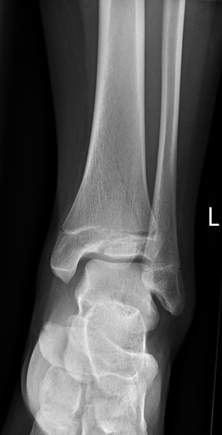
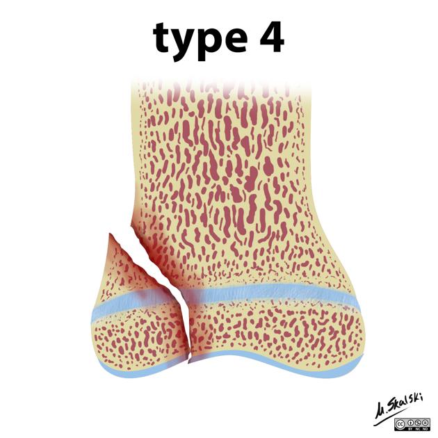
Salter-Harris type IV fractures are relatively uncommon injuries that occur in children. They are intra-articular injuries in which the fracture extends through the epiphysis, across the physis, and through the metaphysis. Salter-Harris fractures are a group of childhood injuries where a fracture involves the physis.
Salter-Harris IV injuries typically have a poor prognosis due to interruption of the proliferative and reserve cartilage zones often leading to altered joint mechanics and functional impairment and as such orthopedic evaluation and subsequent operative intervention are often required 1,2.
Epidemiology
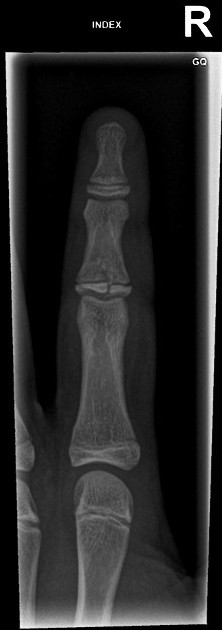
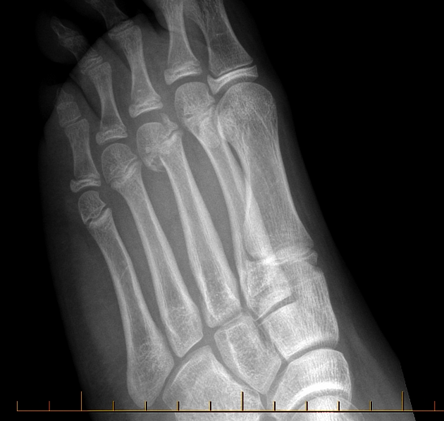
Approximately 10-12% of all physeal fractures will be a Salter-Harris type IV fracture. Salter-Harris type IV injuries will often follow typical location patterns and most commonly involve the distal radius, phalanges, and distal tibia.
Radiographic features
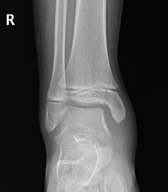
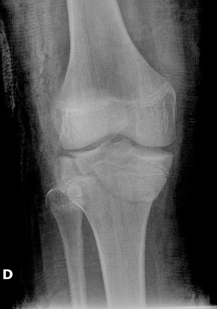
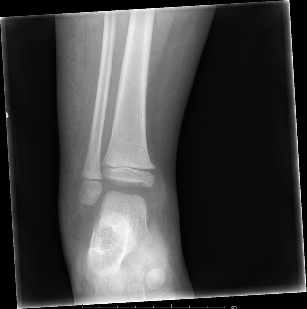
Plain radiograph
lucent fracture line extending through the metaphysis, across physis, and into the epiphysis
angulation, displacement, and rotation may occur
adjacent soft tissue swelling and joint effusion may be noted
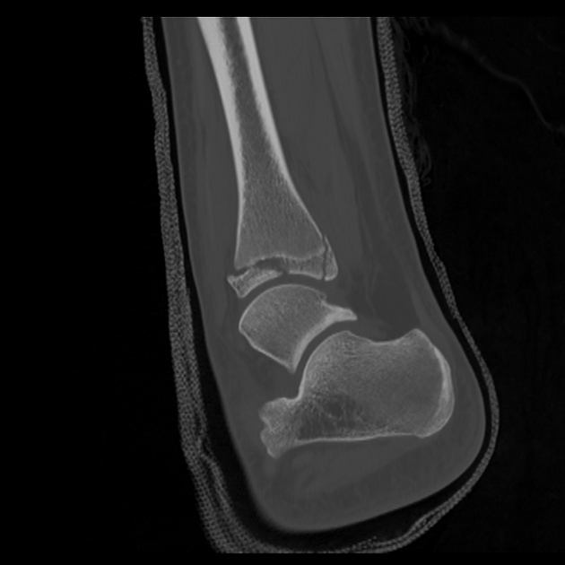
CT
CT may be helpful to further assess the nature of the fracture. This is particularly helpful in the distal tibia when the plain film can under-estimate the complexity and severity of a distal tibial injury.
CT imaging has a role in evaluating the degree of displacement and anatomic extent of Salter-Harris type IV fractures and can subsequently guide operative intervention
CT imaging can also be incorporated to evaluate focal osseous bridging across the physis during the healing process (most common in Salter-Harris IV and V injuries) 3
Quiz questions
References
- 1. Levine R, Thomas A, Nezwek T, Waseem M. Salter-Harris Fracture. 2023. - Pubmed
- 2. Cepela D, Tartaglione J, Dooley T, Patel P. Classifications In Brief: Salter-Harris Classification of Pediatric Physeal Fractures. Clin Orthop Relat Res. 2016;474(11):2531-7. doi:10.1007/s11999-016-4891-3 - Pubmed
- 3. Nguyen J, Markhardt B, Merrow A, Dwek J. Imaging of Pediatric Growth Plate Disturbances. Radiographics. 2017;37(6):1791-812. doi:10.1148/rg.2017170029 - Pubmed
Incoming Links
Related articles: Fractures
-
fracture
- terminology
- fracture location
- diaphyseal fracture
- metaphyseal fracture
- physeal fracture
- epiphyseal fracture
- fracture types[+][+]
- avulsion fracture
- articular surface injuries
- complete fracture
- incomplete fracture
- infraction
- compound fracture
- pathological fracture
- stress fracture
- fracture displacement[+][+]
- fracture location
- fracture healing[+][+]
- skull fractures[+][+]
-
facial fractures[+][+]
- fractures involving a single facial buttress
- alveolar process fractures
- frontal sinus fracture
- isolated zygomatic arch fractures
- mandibular fracture
- nasal bone fracture
- orbital blow-out fracture
- paranasal sinus fractures
- complex fractures
- dental fractures
- fractures involving a single facial buttress
-
spinal fractures[+][+]
- classification (AO Spine classification systems)
-
cervical spine fracture classification systems
- AO classification of upper cervical injuries
- AO classification of subaxial injuries
- Anderson and D'Alonzo classification (odontoid fracture)
- Roy-Camille classification (odontoid process fracture)
- Gehweiler classifcation (atlas fractures)
- Levine and Edwards classification (hangman fracture)
- Allen and Ferguson classification (subaxial spine injuries)
- subaxial cervical spine injury classification (SLIC)
- thoracolumbar spinal fracture classification systems
- three column concept of spinal fractures (Denis classification)
- classification of sacral fractures
-
cervical spine fracture classification systems
- spinal fractures by region
- spinal fracture types
- classification (AO Spine classification systems)
- rib fractures[+][+]
- sternal fractures
-
upper limb fractures[+][+]
- classification
- Rockwood classification (acromioclavicular joint injury)
- AO classification (clavicle fracture)
- Neer classification (clavicle fracture)
- Neer classification (proximal humeral fracture)
- AO classification (proximal humeral fracture)
- AO/OTA classification of distal humeral fractures
- Milch classification (lateral humeral condyle fracture)
- Weiss classification (lateral humeral condyle fracture)
- Bado classification of Monteggia fracture-dislocations (radius-ulna)
- Mason classification (radial head fracture)
- Frykman classification (distal radial fracture)
- Mayo classification (scaphoid fracture)
- Hintermann classification (gamekeeper's thumb)
- Eaton classification (volar plate avulsion injury)
- Keifhaber-Stern classification (volar plate avulsion injury)
- upper limb fractures by region
- shoulder
- clavicular fracture
-
scapular fracture
- acromion fracture
- coracoid process fracture
- glenoid fracture
- humeral head fracture
- proximal humeral fracture
- humeral neck fracture
- arm
- elbow
- forearm
- wrist
-
carpal bones
- scaphoid fracture
- lunate fracture
- capitate fracture
- triquetral fracture
- pisiform fracture
- hamate fracture
- trapezoid fracture
- trapezium fracture
- hand
- shoulder
- classification
- lower limb fractures[+][+]
- classification by region
- pelvic fractures
- hip fractures
- Pipkin classification (femoral head fracture)
- Garden classification (hip fracture)
- American Academy of Orthopedic Surgeons classification (periprosthetic hip fracture)
- Cooke and Newman classification (periprosthetic hip fracture)
- Johansson classification (periprosthetic hip fracture)
- Vancouver classification (periprosthetic hip fracture)
- femoral
- knee
- Schatzker classification (tibial plateau fracture)
- AO classification of distal femur fractures
- Meyers and McKeevers classification (anterior cruciate ligament avulsion fracture)
- tibia/fibula
- Watson-Jones classification (tibial tuberosity avulsion fracture)
- ankle
- foot
- Berndt and Harty classification (osteochondral lesions of the talus)
- Sanders CT classification (calcaneal fracture)
- Hawkins classification (talar neck fracture)
- Myerson classification (Lisfranc injury)
- Nunley-Vertullo classification (Lisfranc injury)
- pelvis and lower limb fractures by region
- pelvic fracture
- sacral fracture
- coccygeal fracture
-
hip
- acetabular fracture
- femoral head fracture
-
femoral neck fracture
- subcapital fracture
- transcervical fracture
- basicervical fracture
-
trochanteric fracture
- pertrochanteric fracture
- intertrochanteric fracture
- subtrochanteric fracture
- femur
- mid-shaft fracture
- bisphosphonate-related fracture
- distal femoral fracture
- knee
- avulsion fractures
- Segond fracture
- reverse Segond fracture
- anterior cruciate ligament avulsion fracture
- posterior cruciate ligament avulsion fracture
- arcuate complex avulsion fracture (arcuate sign)
- biceps femoris avulsion fracture
- iliotibial band avulsion fracture
- semimembranosus tendon avulsion fracture
- Stieda fracture (MCL avulsion fracture)
- patellar fracture
- tibial plateau fracture
- avulsion fractures
- leg
- tibial tuberosity avulsion fracture
- tibial shaft fracture
- fibular shaft fracture
- Maisonneuve fracture
- ankle
- foot
- tarsal bones
- metatarsal bones
- phalanges
- classification by region
- terminology


 Unable to process the form. Check for errors and try again.