Proximal humeral fracture
Proximal humeral fractures are common upper extremity fractures, particularly in older patients, and can result in significant disability.
On this page:
Epidemiology
Proximal humeral fractures represent around 5% of all fractures ?. They are most common in older populations and especially in those who are osteoporotic. As with other injuries, there is a bimodal distribution with a small peak amongst the young.
The majority of proximal humeral fractures occur in the elderly (mean age 65 years) with ~70% occurring in women, presumably due to the greater incidence of osteoporosis 1. Most of these (90%) occur at home due to a fall, and in most cases they are an isolated injury 1.
Clinical presentation
Many older patients present following a relatively innocuous fall. Younger patients usually present following a high-trauma incident, e.g. a motor accident or fall from height. However, patients may present following a seizure, electrical shock or following direct trauma.
Pathology
Proximal humeral fractures usually result from a fall on an outstretched arm. Indirect forces transmitted through the proximal humerus and shoulder are the cause of most fractures. These forces may be compressive, tension, torsion or bending.
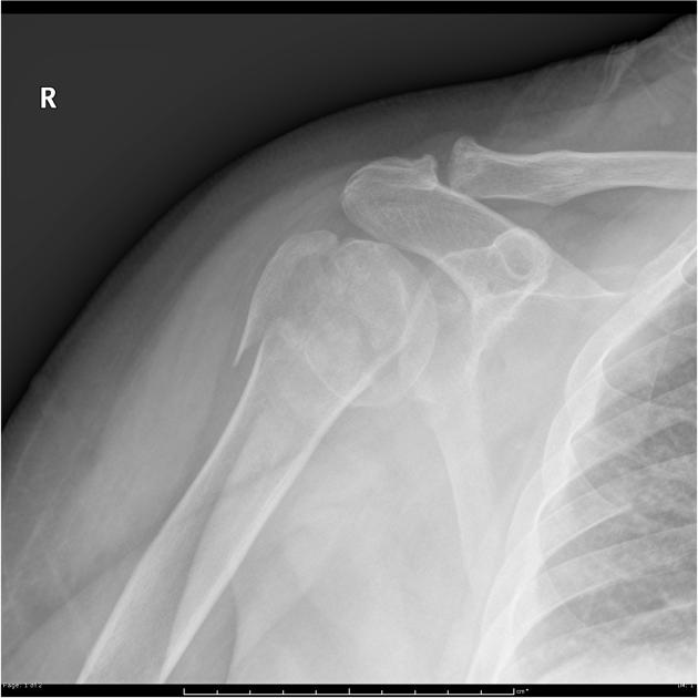
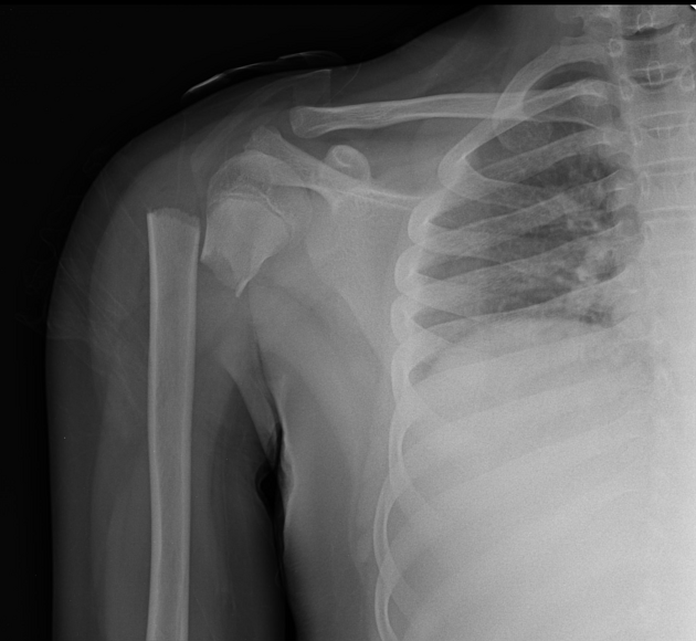
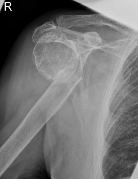
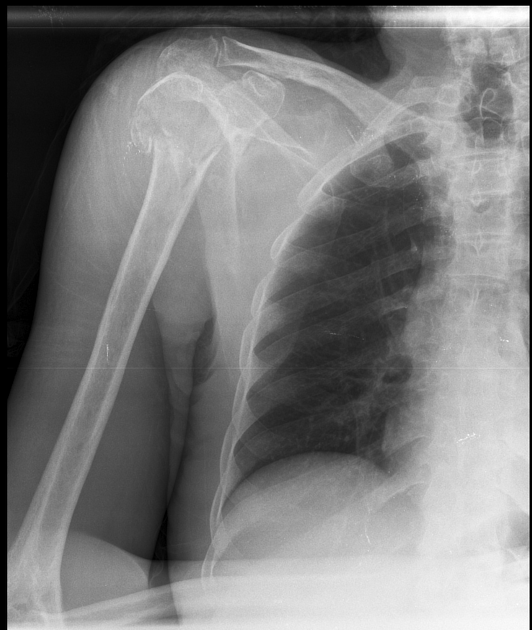
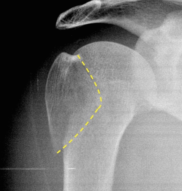
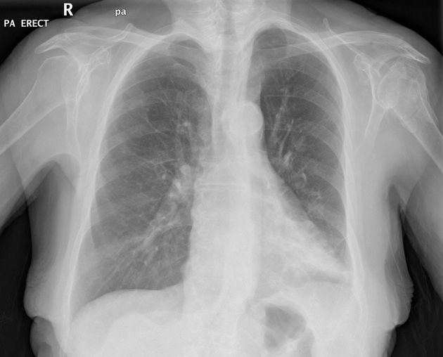
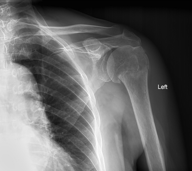
Radiographic features
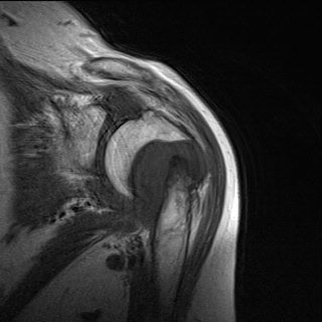
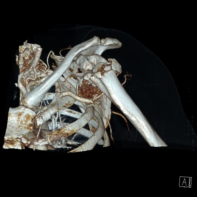
Plain films are usually sufficient to characterize proximal humeral fractures, and thus to determine management. CT can be useful if adequate views cannot be obtained, if fractures are unusual or if other fractures (e.g. glenoid) are present 2. Additionally, CT (and especially 3D surface shaded reconstructions) has been shown to improve interobserver agreement on classification of proximal humeral fractures 4.
Regardless of the imaging performed, the number of displaced fragments should be assessed, to enable appropriate classification of the fracture (Neer classification or AO classification are most commonly used).
Radiograph
The fracture is usually evident as a lucency and cortical breach with variable degrees of angulation, impaction and displacement.
Reporting checklist
In addition to reporting the presence of a fracture, it is important to assess and comment on a number of other features.
- fracture: it should be noted that in most instances a description of the fracture rather than a specific classification is sufficient, but the features required to classify the fracture should be included
- location of fracture lines
- displacement and angulation of each part (>1 cm and >45 degrees respectively is particularly important in the Neer classification)
- presence of involvement of the articular surface
- associated injuries
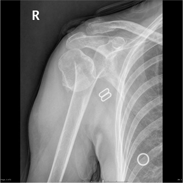
Treatment and prognosis
Management depends not only on the type of fracture but also importantly on the functional status and living situation of the patient. For example, someone who lives alone may not be able to do so without the use of one arm.
Having said this, in almost all cases undisplaced fractures are treated conservatively, whereas operative open reduction and internal fixation (with a variety of intramedullary nails, plates and screws and K-wires) is reserved for displaced fractures 1. Hemi-arthroplasty is also an option especially for three and four-part fractures, where the risk of malunion and avascular necrosis are high 1.
In general, the prognosis is good with the majority of fractures healing well with little functional loss. Poor prognostic factors include 1:
- older age of patient
- displaced fracture
- three and four-part fractures (see Neer classification)
- type C fractures (AO classification)
Related Radiopaedia articles
Fractures
-
fracture
- terminology
- fracture location
- diaphyseal fracture
- metaphyseal fracture
- physeal fracture
- epiphyseal fracture
- fracture types
- avulsion fracture
- articular surface injuries
- complete fracture
- incomplete fracture
- infraction
- compound fracture
- pathological fracture
- stress fracture
- fracture displacement
- fracture location
- fracture healing
- skull fractures
-
facial fractures
- fractures involving a single facial buttress
- alveolar process fractures
- frontal sinus fracture
- isolated zygomatic arch fractures
- mandibular fracture
- nasal bone fracture
- orbital blow-out fracture
- paranasal sinus fractures
- complex fractures
- dental fractures
- fractures involving a single facial buttress
-
spinal fractures
- classification (AO Spine classification systems)
-
cervical spine fracture classification systems
- AO classification of upper cervical injuries
- AO classification of subaxial injuries
- Anderson and D'Alonzo classification (odontoid fracture)
- Levine and Edwards classification (hangman fracture)
- Roy-Camille classification (odontoid process fracture )
- Allen and Ferguson classification (subaxial spine injuries)
- subaxial cervical spine injury classification (SLIC)
- thoracolumbar spinal fracture classification systems
- three column concept of spinal fractures (Denis classification)
- classification of sacral fractures
-
cervical spine fracture classification systems
- spinal fractures by region
- spinal fracture types
- classification (AO Spine classification systems)
- rib fractures
- sternal fractures
-
upper limb fractures
- classification
- Rockwood classification (acromioclavicular joint injury)
- AO classification (clavicle fracture)
- Neer classification (clavicle fracture)
- Neer classification (proximal humeral fracture)
- AO classification (proximal humeral fracture)
- AO/OTA classification of distal humeral fractures
- Milch classification (lateral humeral condyle fracture)
- Weiss classification (lateral humeral condyle fracture)
- Bado classification of Monteggia fracture-dislocations (radius-ulna)
- Mason classification (radial head fracture)
- Frykman classification (distal radial fracture)
- Mayo classification (scaphoid fracture)
- Hintermann classification (gamekeeper's thumb)
- Eaton classification (volar plate avulsion injury)
- Keifhaber-Stern classification (volar plate avulsion injury)
- upper limb fractures by region
- shoulder
- clavicular fracture
-
scapular fracture
- acromion fracture
- coracoid process fracture
- glenoid fracture
- humeral head fracture
- proximal humeral fracture
- humeral neck fracture
- arm
- elbow
- forearm
- wrist
-
carpal bones
- scaphoid fracture
- lunate fracture
- capitate fracture
- triquetral fracture
- pisiform fracture
- hamate fracture
- trapezoid fracture
- trapezium fracture
- hand
- shoulder
- classification
- lower limb fractures
- classification by region
- pelvis
- hip
- Pipkin classification (femoral head fracture)
- Garden classification (hip fracture)
- American Academy of Orthopedic Surgeons classification (periprosthetic hip fracture)
- Cooke and Newman classification (periprosthetic hip fracture)
- Johansson classification (periprosthetic hip fracture)
- Vancouver classification (periprosthetic hip fracture)
- femoral
- knee
- Schatzker classification (tibial plateau fracture)
- Meyers and McKeevers classification (anterior cruciate ligament avulsion fracture)
- tibia/fibula
- Watson-Jones classification (tibial tuberosity avulsion fracture)
- ankle
- foot
- Berndt and Harty classification (osteochondral lesions of the talus)
- Sanders CT classification (calcaneal fracture)
- Hawkins classification (talar neck fracture)
- Myerson classification (Lisfranc injury)
- Nunley-Vertullo classification (Lisfranc injury)
- pelvis and lower limb fractures by region
- pelvic fracture
- sacral fracture
- coccygeal fracture
-
hip
- acetabular fracture
- femoral head fracture
-
femoral neck fracture
- subcapital fracture
- transcervical fracture
- basicervical fracture
-
trochanteric fracture
- pertrochanteric fracture
- intertrochanteric fracture
- subtrochanteric fracture
- thigh
- mid-shaft fracture
- bisphosphonate-related fracture
- knee
- avulsion fractures
- Segond fracture
- reverse Segond fracture
- anterior cruciate ligament avulsion fracture
- posterior cruciate ligament avulsion fracture
- arcuate complex avulsion fracture (arcuate sign)
- biceps femoris avulsion fracture
- iliotibial band avulsion fracture
- semimembranosus tendon avulsion fracture
- Stieda fracture (MCL avulsion fracture)
- patellar fracture
- tibial plateau fracture
- avulsion fractures
- leg
- tibial tuberosity avulsion fracture
- tibial shaft fracture
- fibular shaft fracture
- Maisonneuve fracture
- ankle
- foot
- tarsal bones
- metatarsal bones
- phalanges
- classification by region
- terminology