
Subchondral insufficiency fracture
Citation, DOI, disclosures and article data
At the time the article was created Yuranga Weerakkody had no recorded disclosures.
View Yuranga Weerakkody's current disclosuresAt the time the article was last revised Joshua Yap had no financial relationships to ineligible companies to disclose.
View Joshua Yap's current disclosures- Subchondral insufficiency fractures
Subchondral insufficiency fracture refers to a type of stress fracture that occurs below the chondral surface on a weight-bearing surface of a bone due to mechanic failure of subchondral cancellous bone.
On this page:
Epidemiology
Risk factors
obesity
abnormal loading through the joint
overuse
Pathology
They tend to occur when normal physiological forces are repeatedly applied to an area of bone. Callus formation occurs along with non-mineralized osteoid and the absence of bone infarction.
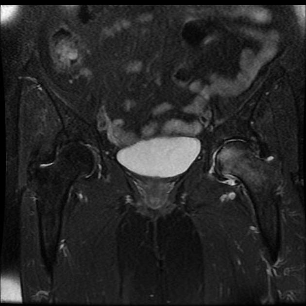
Location
Typical sites include:
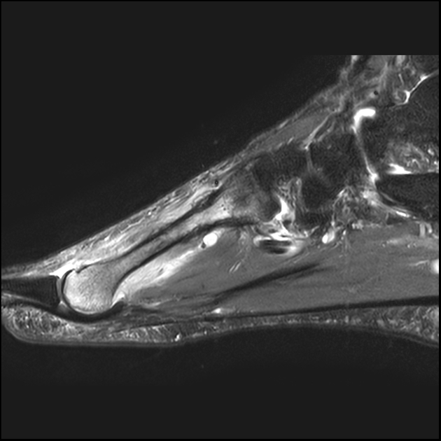
femoral head
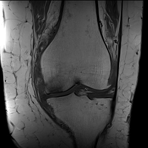
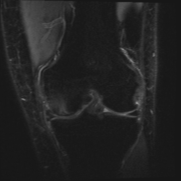
femoral condyles
tibial plateau
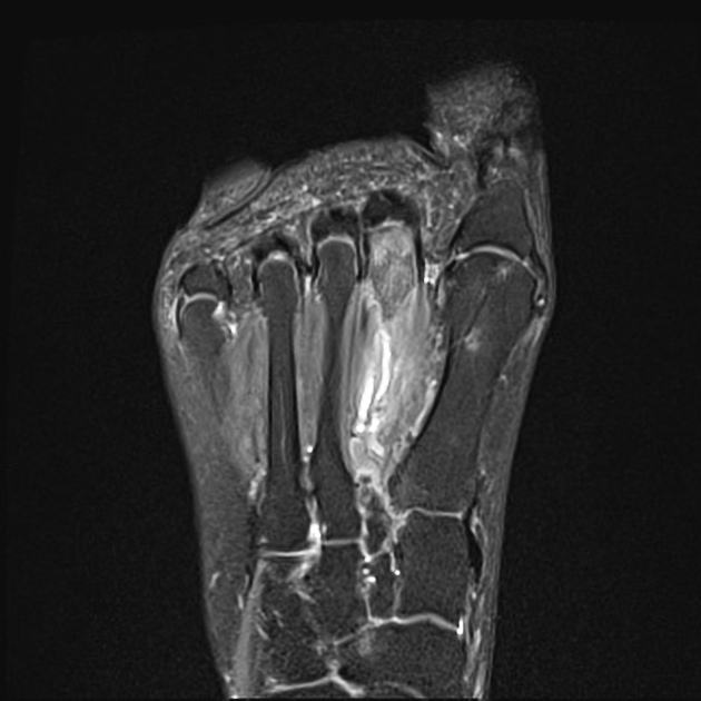
metatarsal head
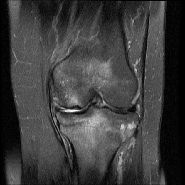
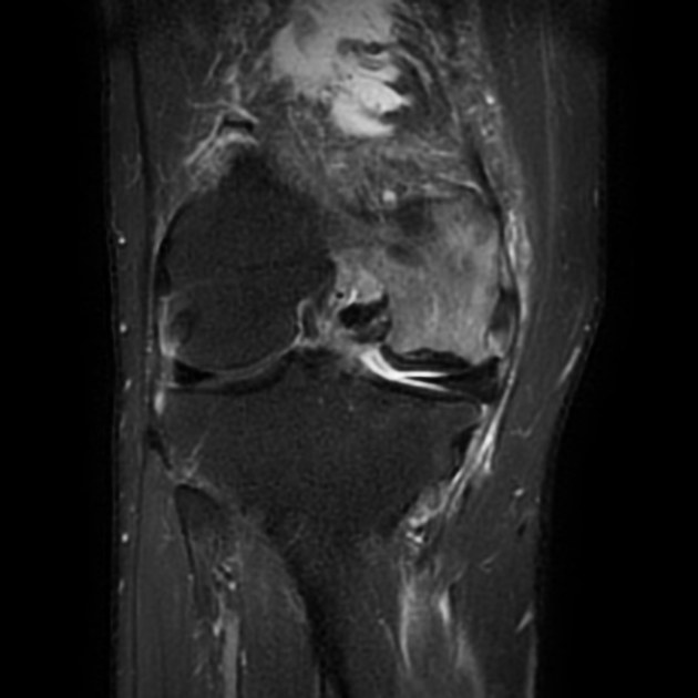
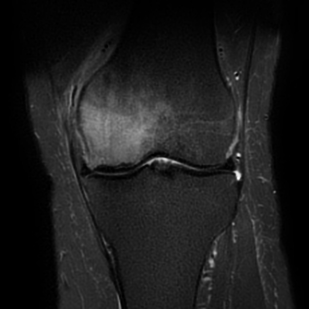
Radiographic features
Plain radiograph / CT
Radiographs are normal preceding the development of callus formation or collapse of the articular surface. Linear or patchy subchondral sclerosis may be present representing cancellous fracture.
MRI
-
T1: there may be a characteristically low signal intensity band through the affected region
parallels the subchondral bone plate (whereas osteonecrosis is curvilinear)
preservation of the articular cartilage (unlike an osteochondral defect)
T2: florid marrow edema
Treatment and prognosis
Complications
secondary osteonecrosis
osteonecrosis with cavitation (crescent sign)
articular collapse
History and etymology
It has been described in many regions but the term "subchondral insufficiency fracture of the femoral head" was coined by Bangil et al. in 1996 7.
Differential diagnosis
The differential diagnosis of subchondral marrow edema includes:
See also
References
- 1. Yamamoto T. Subchondral insufficiency fractures of the femoral head. (2012) Clinics in orthopedic surgery. 4 (3): 173-80. doi:10.4055/cios.2012.4.3.173 - Pubmed
- 2. Yamamoto T, Takabatake K, Iwamoto Y. Subchondral insufficiency fracture of the femoral head resulting in rapid destruction of the hip joint: a sequential radiographic study. (2002) AJR. American journal of roentgenology. 178 (2): 435-7. doi:10.2214/ajr.178.2.1780435 - Pubmed
- 3. An VV, Broek MVD, Oussedik S. Subchondral Insufficiency Fracture in the Lateral Compartment of the Knee in a 64-Year-Old Marathon Runner. (2017) Knee surgery & related research. 29 (4): 325-328. doi:10.5792/ksrr.17.049 - Pubmed
- 4. Jose J, Pasquotti G, Smith MK, Gupta A, Lesniak BP, Kaplan LD. Subchondral insufficiency fractures of the knee: review of imaging findings. (2015) Acta radiologica (Stockholm, Sweden : 1987). 56 (6): 714-9. doi:10.1177/0284185114535132 - Pubmed
- 5. Vincent VG An, Mathias van den Broek, and Sam Oussedik. Subchondral Insufficiency Fracture in the Lateral Compartment of the Knee in a 64-Year-Old Marathon Runner. (2017) Knee Surgery and Related Research. 29 (4): 325. doi:10.5792/ksrr.17.049 - Pubmed
- 6. William Palmer, Laura Bancroft, Fiona Bonar, Jung-Ah Choi, Anne Cotten, James F. Griffith, Philip Robinson, Christian W.A. Pfirrmann. Glossary of terms for musculoskeletal radiology. (2020) Skeletal Radiology. doi:10.1007/s00256-020-03465-1 - Pubmed
- 7. Ghate SD, Samant A. Subchondral Insufficiency Fracture of Femoral head: Uncommon cause of Hip pain in Elderly. (2012) Journal of orthopaedic case reports. 2 (2): 7-9. Pubmed
Incoming Links
- Subchondral insufficiency fracture of the knee
- Foot radiograph (checklist)
- Osteonecrosis of the femoral head
- Femoroacetabular impingement syndrome
- Osteoarthritis of the hip
- Subchondral fracture
- Osteochondral defect
- Acetabular dysplasia
- Osteomeniscal impact oedema
- Differential diagnosis for metatarsal region pain
- Freiberg disease
- Subchondral insuffiency fracture of the acetabulum
- Subchondral insufficiency fracture of the knee
- Subchondral insufficiency fracture - 2nd metatarsal head
- Subchondral insufficiency fracture of the knee
- Subchondral insufficiency fracture - left femoral head
- Subchondral insufficiency fracture - femoral head
- Subchondral insufficiency fracture - femoral head
- Subchondral insufficiency fracture of talus
- Subchondral insufficiency fracture - femoral head
- Tibial plateau insufficiency fracture
- Spontaneous osteonecrosis of the knee
- Metatarsal fatigue and insufficiency (stress) fractures
- Subchondral insufficiency fracture post lateral meniscectomy
- Subchondral insufficiency fracture - knee
- Subchondral insufficiency fracture - metatarsal head
Related articles: Fractures
-
fracture
- terminology
- fracture location[+][+]
- diaphyseal fracture
- metaphyseal fracture
- physeal fracture
- epiphyseal fracture
- fracture types
- avulsion fracture
- articular surface injuries
- bone contusion
- chondral fracture
-
subchondral fracture
- subchondral insufficiency fracture
- osteochondral fracture
- complete fracture[+][+]
- incomplete fracture[+][+]
- infraction
- compound fracture[+][+]
- pathological fracture
- stress fracture[+][+]
- fracture displacement[+][+]
- fracture location[+][+]
- fracture healing[+][+]
- skull fractures[+][+]
-
facial fractures[+][+]
- fractures involving a single facial buttress
- alveolar process fractures
- frontal sinus fracture
- isolated zygomatic arch fractures
- mandibular fracture
- nasal bone fracture
- orbital blow-out fracture
- paranasal sinus fractures
- complex fractures
- dental fractures
- fractures involving a single facial buttress
-
spinal fractures[+][+]
- classification (AO Spine classification systems)
-
cervical spine fracture classification systems
- AO classification of upper cervical injuries
- AO classification of subaxial injuries
- Anderson and D'Alonzo classification (odontoid fracture)
- Roy-Camille classification (odontoid process fracture)
- Gehweiler classifcation (atlas fractures)
- Levine and Edwards classification (hangman fracture)
- Allen and Ferguson classification (subaxial spine injuries)
- subaxial cervical spine injury classification (SLIC)
- thoracolumbar spinal fracture classification systems
- three column concept of spinal fractures (Denis classification)
- classification of sacral fractures
-
cervical spine fracture classification systems
- spinal fractures by region
- spinal fracture types
- classification (AO Spine classification systems)
- rib fractures[+][+]
- sternal fractures
-
upper limb fractures[+][+]
- classification
- Rockwood classification (acromioclavicular joint injury)
- AO classification (clavicle fracture)
- Neer classification (clavicle fracture)
- Neer classification (proximal humeral fracture)
- AO classification (proximal humeral fracture)
- AO/OTA classification of distal humeral fractures
- Milch classification (lateral humeral condyle fracture)
- Weiss classification (lateral humeral condyle fracture)
- Bado classification of Monteggia fracture-dislocations (radius-ulna)
- Mason classification (radial head fracture)
- Frykman classification (distal radial fracture)
- Mayo classification (scaphoid fracture)
- Hintermann classification (gamekeeper's thumb)
- Eaton classification (volar plate avulsion injury)
- Keifhaber-Stern classification (volar plate avulsion injury)
- upper limb fractures by region
- shoulder
- clavicular fracture
-
scapular fracture
- acromion fracture
- coracoid process fracture
- glenoid fracture
- humeral head fracture
- proximal humeral fracture
- humeral neck fracture
- arm
- elbow
- forearm
- wrist
-
carpal bones
- scaphoid fracture
- lunate fracture
- capitate fracture
- triquetral fracture
- pisiform fracture
- hamate fracture
- trapezoid fracture
- trapezium fracture
- hand
- shoulder
- classification
- lower limb fractures[+][+]
- classification by region
- pelvic fractures
- hip fractures
- Pipkin classification (femoral head fracture)
- Garden classification (hip fracture)
- American Academy of Orthopedic Surgeons classification (periprosthetic hip fracture)
- Cooke and Newman classification (periprosthetic hip fracture)
- Johansson classification (periprosthetic hip fracture)
- Vancouver classification (periprosthetic hip fracture)
- femoral
- knee
- Schatzker classification (tibial plateau fracture)
- AO classification of distal femur fractures
- Meyers and McKeevers classification (anterior cruciate ligament avulsion fracture)
- tibia/fibula
- Watson-Jones classification (tibial tuberosity avulsion fracture)
- ankle
- foot
- Berndt and Harty classification (osteochondral lesions of the talus)
- Sanders CT classification (calcaneal fracture)
- Hawkins classification (talar neck fracture)
- Myerson classification (Lisfranc injury)
- Nunley-Vertullo classification (Lisfranc injury)
- pelvis and lower limb fractures by region
- pelvic fracture
- sacral fracture
- coccygeal fracture
-
hip
- acetabular fracture
- femoral head fracture
-
femoral neck fracture
- subcapital fracture
- transcervical fracture
- basicervical fracture
-
trochanteric fracture
- pertrochanteric fracture
- intertrochanteric fracture
- subtrochanteric fracture
- femur
- mid-shaft fracture
- bisphosphonate-related fracture
- distal femoral fracture
- knee
- avulsion fractures
- Segond fracture
- reverse Segond fracture
- anterior cruciate ligament avulsion fracture
- posterior cruciate ligament avulsion fracture
- arcuate complex avulsion fracture (arcuate sign)
- biceps femoris avulsion fracture
- iliotibial band avulsion fracture
- semimembranosus tendon avulsion fracture
- Stieda fracture (MCL avulsion fracture)
- patellar fracture
- tibial plateau fracture
- avulsion fractures
- leg
- tibial tuberosity avulsion fracture
- tibial shaft fracture
- fibular shaft fracture
- Maisonneuve fracture
- ankle
- foot
- tarsal bones
- metatarsal bones
- phalanges
- classification by region
- terminology


 Unable to process the form. Check for errors and try again.