
Arachnoid mater
Citation, DOI, disclosures and article data
At the time the article was created Julian Maingard had no recorded disclosures.
View Julian Maingard's current disclosuresAt the time the article was last revised Daniel J Bell had no financial relationships to ineligible companies to disclose.
View Daniel J Bell's current disclosuresThe arachnoid mater forms the middle layer of the meninges and together with the pia mater is sometimes referred to as the leptomeninges.
On this page:
Gross anatomy
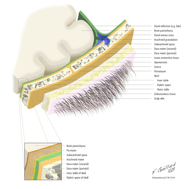
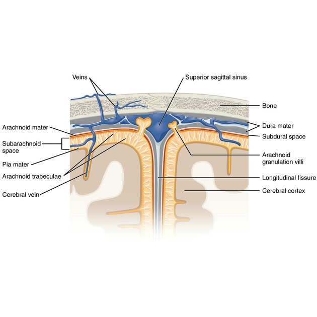
The arachnoid mater is a membrane that comes into direct contact with the dura mater and is separated from the pia mater by a CSF-filled space known as the subarachnoid space 1.
The arachnoid mater loosely surrounds the entire brain and is easily separable from the dura except where it is adherent to the adventitia of the internal carotid and vertebral arteries at the point in which they enter the subarachnoid space 1. It extends to the superior surface of the pituitary fossa but does not envelop the pituitary gland itself. At the level of the foramen magnum, the cerebral arachnoid mater is continuous with the spinal arachnoid mater. The arachnoid mater also exits the cranium as part of the optic nerve-sheath complex 2.
It does not enter the sulci or fissures except at the great longitudinal fissure between the cerebral hemispheres. It is loosely associated with the pia mater via a network of connective tissue that crosses the subarachnoid space known as arachnoid trabeculae, arachnoid septa and arachnoid membranes (some of which are named) 2,3.
Outpouchings of the arachnoid mater that pierce the dura in the venous dural sinuses are known as arachnoid granulations and are involved in CSF reabsorption 2.
History and etymology
The term arachnoid is derived from the Greek word "arachne" meaning "spider" on account of the delicate web-like appearance of the arachnoid membranes, trabeculae and septae that traverse the subarachnoid space from the arachnoid mater to the pia mater.
Related pathology
Quiz questions
References
- 1. Richard Lee Drake, Henry Gray, Wayne Vogl et al. Gray's Anatomy for Students. (2005) ISBN: 9780443071683 - Google Books
- 2. Adeeb N, Deep A, Griessenauer C et al. The Intracranial Arachnoid Mater : A Comprehensive Review of Its History, Anatomy, Imaging, and Pathology. Childs Nerv Syst. 2013;29(1):17-33. doi:10.1007/s00381-012-1910-x - Pubmed
- 3. Matsuno H, Rhoton A, Peace D. Microsurgical Anatomy of the Posterior Fossa Cisterns. Neurosurgery. 1988;23(1):58-80. doi:10.1227/00006123-198807000-00012 - Pubmed
Incoming Links
- Subdural space
- Intralaminar dural haemorrhage
- Subarachnoid space
- Chondrosarcoma of the skull base
- Tuberculous meningitis
- Leptomeninges
- Blood supply of the meninges
- Subarachnoid cisterns
- Central artery of the retina
- Dura mater
- Spinal dura mater
- Cranial meninges
- Subdural empyema
- Leptomeningitis
- Arachnoid membranes
- Meninx primitiva
- Accessory nerve
- Pachymeningeal enhancement
- Meningeal enhancement
- Subdural haemorrhage (summary)
Related articles: Anatomy: Brain
-
brain
- grey matter
- white matter
-
cerebrum
-
cerebral hemisphere (telencephalon)
- cerebral lobes and gyri
- frontal lobe
- parietal lobe
-
occipital lobe
- occipital pole
- lingual gyrus
- fusiform gyrus (Brodmann area 37)
- calcarine (visual) cortex
- cuneus
- temporal lobe
- basal forebrain
- limbic system
- insula
-
cerebral sulci and fissures (A-Z)
- calcarine fissure
- callosal sulcus
- central (Rolandic) sulcus
- cingulate sulcus
- collateral sulcus
- inferior frontal sulcus
- inferior occipital sulcus
- inferior temporal sulcus
- interhemispheric fissure
- intraparietal sulcus
- lateral (Sylvian) sulcus
- lateral occipital sulcus
- marginal sulcus
- occipitotemporal sulcus
- olfactory sulcus
- paracentral sulcus
- paraolfactory sulcus
- parieto-occipital fissure
- posterior parolfactory sulcus
- precentral sulcus
- preoccipital notch
- postcentral sulcus
- rhinal sulcus
- rostral sulcus
- subparietal sulcus
- superior frontal sulcus
- superior occipital sulcus
- superior temporal sulcus
- cortical histology
- cerebral lobes and gyri
- white matter tracts
- deep grey matter
-
pituitary gland
- posterior pituitary and stalk (part of diencephalon)
- anterior pituitary
- inferior hypophyseal arterial circle
- diencephalon
-
cerebral hemisphere (telencephalon)
-
brainstem
- midbrain (mesencephalon)
- pons (part of metencephalon)
- medulla oblongata (myelencephalon)
- white matter
-
grey matter
- non-cranial nerve
-
cranial nerve nuclei
- oculomotor nucleus
- Edinger-Westphal nucleus
- trochlear nucleus
- motor nucleus of CN V
- mesencephalic nucleus of CN V
- main sensory nucleus of CN V
- spinal nucleus of CN V
- abducent nucleus
- facial nucleus
- superior salivatory nucleus
- cochlear nuclei
- vestibular nuclei
- inferior salivatory nucleus
- solitary tract nucleus
- ambiguus nucleus
- dorsal vagal motor nucleus
- hypoglossal nucleus
-
cerebellum (part of metencephalon)
- vermis
- cerebellar hemisphere
- cerebellar peduncles
- cranial meninges (meninx primitiva)
- CSF spaces
-
cranial nerves (mnemonic)
- olfactory nerve (CN I)
- optic nerve (CN II)
- oculomotor nerve (CN III)
- trochlear nerve (CN IV)
- trigeminal nerve (CN V) (mnemonic)
- abducens nerve (CN VI)
- facial nerve (CN VII) (segments mnemonic | branches mnemonic)
-
vestibulocochlear nerve (CN VIII)
- vestibular ganglion (Scarpa's ganglion)
- glossopharyngeal nerve (CN IX)
- vagus nerve (CN X)
- spinal accessory nerve (CN XI)
- hypoglossal nerve (CN XII)
- functional neuroanatomy
- CNS development
- cerebral vascular supply
- arteries
- vascular territories
-
circle of Willis
- internal carotid artery (ICA) (segments)
- vertebral artery
-
normal variants
- intracranial arterial fenestration
- internal carotid artery (ICA)
- anterior cerebral artery (ACA)
- middle cerebral artery (MCA)
- posterior cerebral artery (PCA)
- basilar artery
- persistent carotid-vertebrobasilar artery anastomoses (mnemonic)
- vertebral artery
- ophthalmic artery
-
cerebral venous system
-
dural venous sinuses
- basilar venous plexus
- cavernous sinus (mnemonic)
- clival diploic veins
- inferior petro-occipital vein
- inferior petrosal sinus
- inferior sagittal sinus
- intercavernous sinus
- internal carotid artery venous plexus of Rektorzik
- jugular bulb
- marginal sinus
- occipital sinus
- sigmoid sinus
- sphenoparietal sinus
- straight sinus
- superior petrosal sinus
- superior sagittal sinus
- torcula herophili
- transverse sinus
-
cerebral veins
-
superficial veins of the brain
- superior cerebral veins (superficial cerebral veins)
- inferior cerebral veins
- superficial middle cerebral vein
- superior anastomotic vein (of Trolard)
- inferior anastomotic vein (of Labbe)
-
superficial veins of the brain
-
deep veins of the brain
- great cerebral vein (of Galen)
- venous circle of Trolard
- normal variants
-
dural venous sinuses
- arteries
- glymphatic pathway


 Unable to process the form. Check for errors and try again.