
Fetal posterior cerebral artery
Citation, DOI, disclosures and article data
At the time the article was created Frank Gaillard had no recorded disclosures.
View Frank Gaillard's current disclosuresAt the time the article was last revised Henry Knipe had the following disclosures:
- Integral Diagnostics, Shareholder (ongoing)
- Micro-X Ltd, Shareholder (ongoing)
These were assessed during peer review and were determined to not be relevant to the changes that were made.
View Henry Knipe's current disclosures- Fetal posterior comminucating artery
- Fetal origin of posterior cerebral artery
- Fetal posterior cerebral arteries
- Fetal PCOM
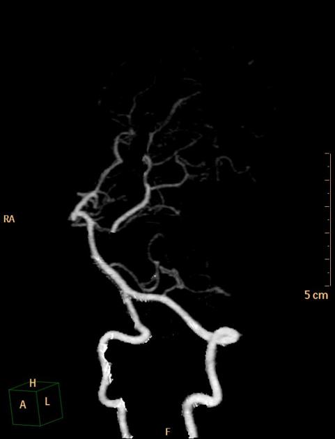
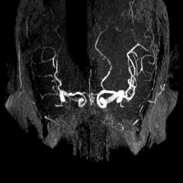
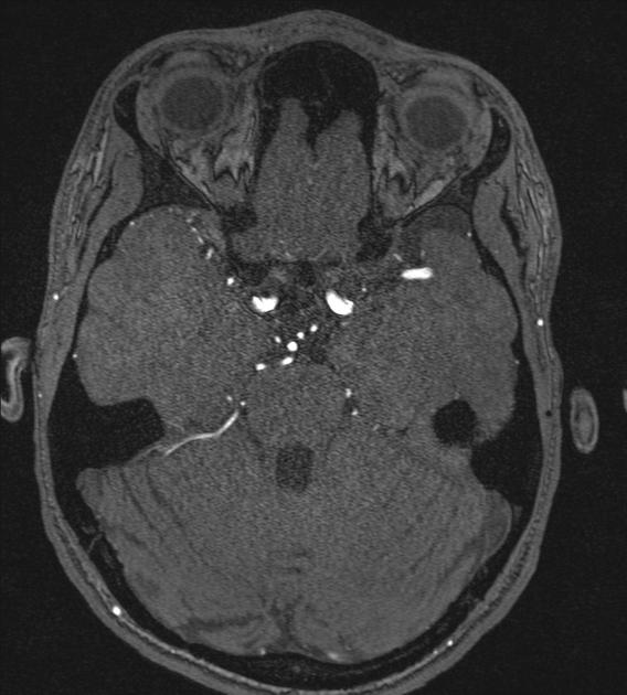
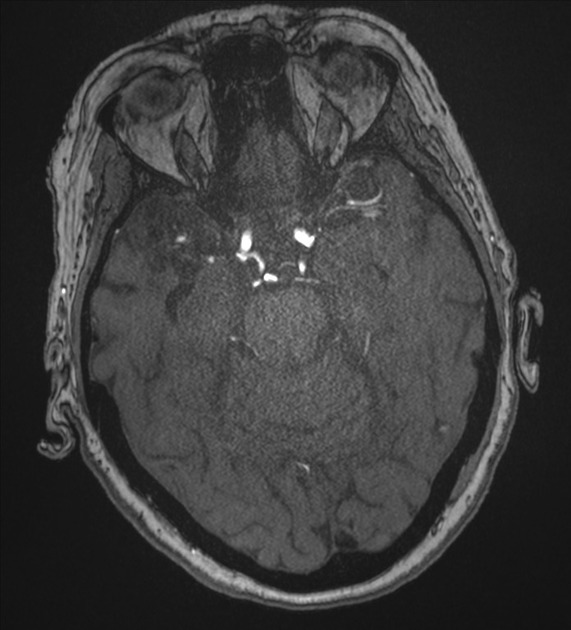
A fetal (origin of the) posterior cerebral artery (fetal PCA), sometimes also referred to less accurately as fetal (origin of the) posterior communicating artery (fetal PCom), is a common variant in the posterior cerebral circulation.
On this page:
Epidemiology
Fetal PCAs occur in ~25% (range 20-30%) of individuals 2.
Gross anatomy
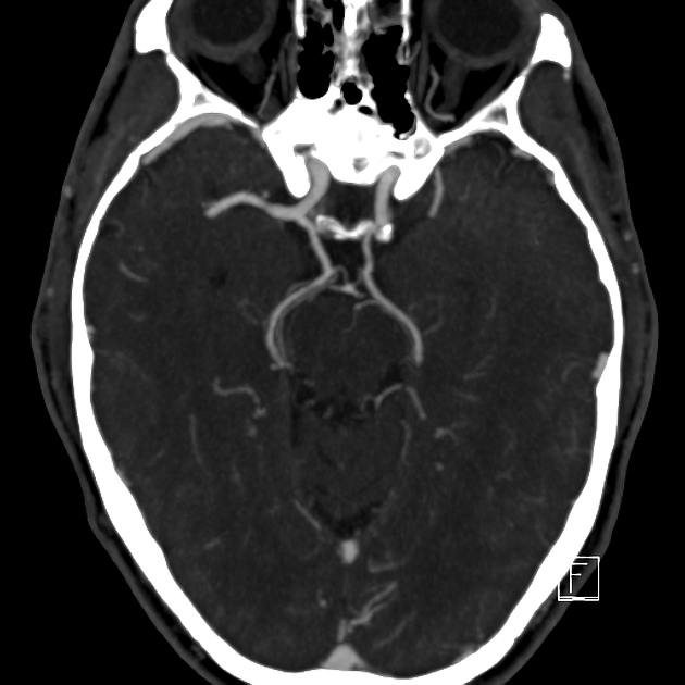
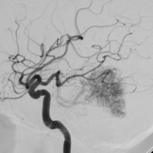
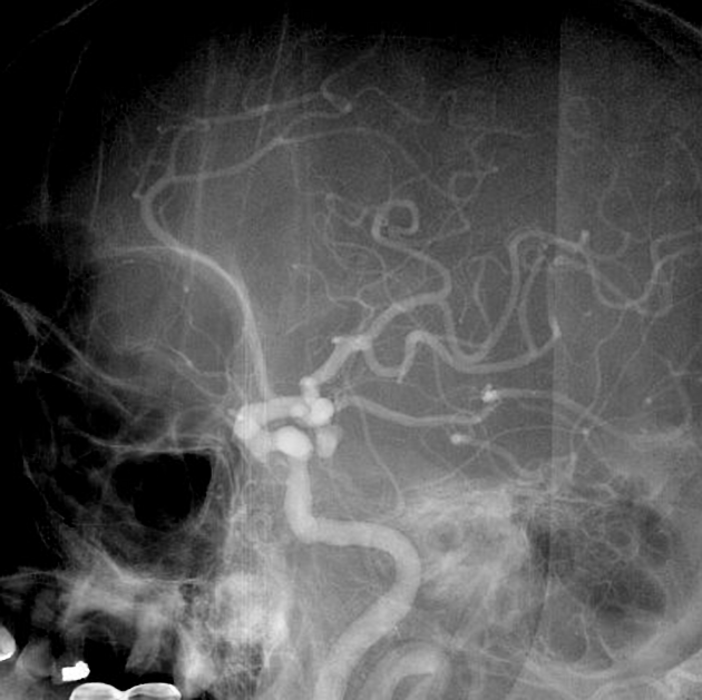
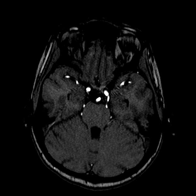
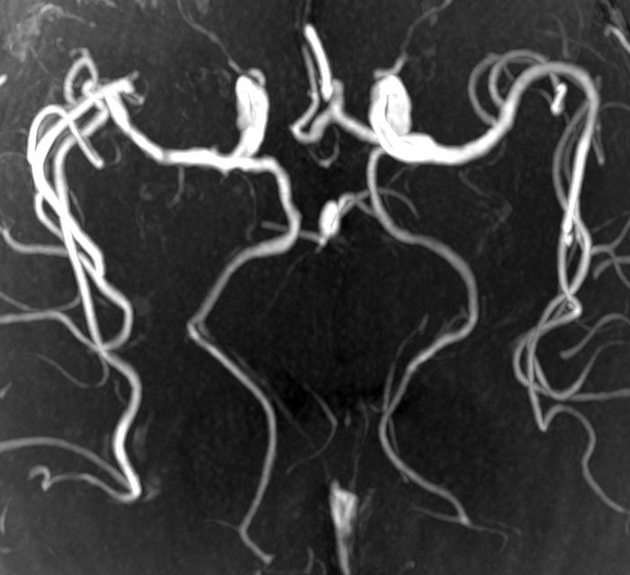
A fetal PCA describes a situation whereby the posterior communicating artery (PCom) is larger than the P1 segment of the posterior cerebral artery (PCA) and, thus, supplies the bulk of the blood to the PCA 4,5. The P1 can be small (hypoplastic) or absent in this setting. When bilateral fetal PCAs are present, the basilar artery will be significantly smaller in caliber than normal ref.
Relations
In cases of non-fetal PCA, the PCom lies superomedial to the oculomotor nerve, whereas, in cases of fetal PCA, it lies superior or superolateral to it ref.
Clinical significance
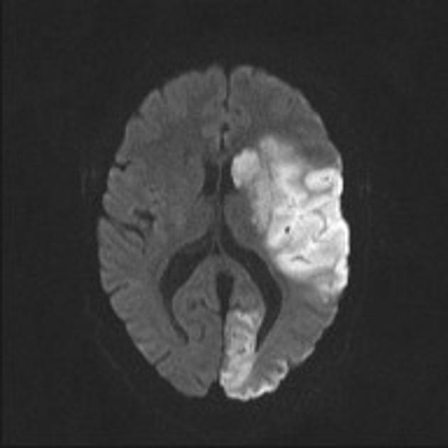
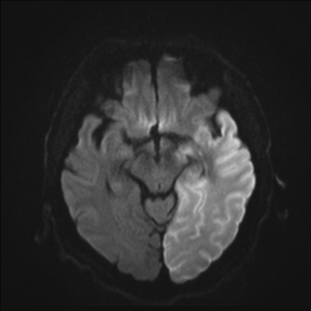
The significance of fetal PCA is in relation to the pattern of ischemic stroke, given that if a fetal PCA is present, the PCA is essentially part of the anterior circulation ref. This becomes relevant, for example, if there is ipsilateral internal carotid artery stenosis. Additionally, a larger PCom with an existing P1 allows for collateral circulation.

References
- 1. Yamamoto Y, Georgiadis A, Chang H, Caplan L. Posterior Cerebral Artery Territory Infarcts in the New England Medical Center Posterior Circulation Registry. Arch Neurol. 1999;56(7):824-32. doi:10.1001/archneur.56.7.824 - Pubmed
- 2. Zampakis P, Panagiotopoulos V, Petsas T, Kalogeropoulou C. Common and Uncommon Intracranial Arterial Anatomic Variations in Multi-Detector Computed Tomography Angiography (MDCTA). What Radiologists Should Be Aware Of. Insights Imaging. 2015;6(1):33-42. doi:10.1007/s13244-014-0381-x - Pubmed
- 3. Lambert SL, Williams FJ, Oganisyan ZZ et-al. Fetal-Type Variants of the Posterior Cerebral Artery and Concurrent Infarction in the Major Arterial Territories of the Cerebral Hemisphere. (2016) Journal of investigative medicine high impact case reports. 4 (3): 2324709616665409. doi:10.1177/2324709616665409 - Pubmed
- 4. Capone S, Shah N, George-St Bernard R. A Fetal-Type Variant Posterior Communicating Artery and Its Clinical Significance. Cureus. 2019;11(7):e5064. doi:10.7759/cureus.5064 - Pubmed
- 5. Mohamed Micdhadhu M, Kho K, Murad M, Looi I. Fetal Posterior Communicating Artery as a Conduit for Concurrent Anterior and Posterior Circulation Infarct: A Case Report. CVNS. 2021;3(4):7-11. doi:10.32896/cvns.v3n4.7-11
Incoming Links
- Fetal posterior cerebral artery
- Aplastic right A1 and bilateral fetal posterior communicating arteries on MRA
- Posterior cerebral artery infarct
- Subarachnoid hemorrhage due to ruptured posterior communicating artery aneurysm
- Persistent primitive trigeminal artery- Saltzman type II
- Twig-like middle cerebral artery
- Bilateral fetal origin posterior cerebral arteries with hypoplastic basilar artery
- Qudrigeminal cistern lipoma and multiple anatomic variations of intracranial arteries
- A1 segment hypoplasia
- Fetal posterior communicating artery with aneurysm
- Ischaemic stroke - dense posterior cerebral artery (PCA) sign
- Internal carotid artery hypoplasia
- Aplasia of the internal carotid artery
- Common variants of the circle of Willis (illustrations)
- Lennox-Gastaut syndrome
- Primary intraventricular hemorrhage: cause unknown
- Primary intraventricular hemorrhage: cause unknown
- Bilateral fetal posterior communicating arteries
- Fetal posterior communicating artery
- Fetal posterior communicating artery
Related articles: Anatomy: Brain
-
brain
- grey matter
- white matter
-
cerebrum
-
cerebral hemisphere (telencephalon)
- cerebral lobes and gyri
- frontal lobe
- parietal lobe
-
occipital lobe
- occipital pole
- lingual gyrus
- fusiform gyrus (Brodmann area 37)
- calcarine (visual) cortex
- cuneus
- temporal lobe
- basal forebrain
- limbic system
- insula
-
cerebral sulci and fissures (A-Z)
- calcarine fissure
- callosal sulcus
- central (Rolandic) sulcus
- cingulate sulcus
- collateral sulcus
- inferior frontal sulcus
- inferior occipital sulcus
- inferior temporal sulcus
- interhemispheric fissure
- intraparietal sulcus
- lateral (Sylvian) sulcus
- lateral occipital sulcus
- marginal sulcus
- occipitotemporal sulcus
- olfactory sulcus
- paracentral sulcus
- paraolfactory sulcus
- parieto-occipital fissure
- posterior parolfactory sulcus
- precentral sulcus
- preoccipital notch
- postcentral sulcus
- rhinal sulcus
- rostral sulcus
- subparietal sulcus
- superior frontal sulcus
- superior occipital sulcus
- superior temporal sulcus
- cortical histology
- cerebral lobes and gyri
- white matter tracts
- deep grey matter
-
pituitary gland
- posterior pituitary and stalk (part of diencephalon)
- anterior pituitary
- inferior hypophyseal arterial circle
- diencephalon
-
cerebral hemisphere (telencephalon)
-
brainstem
- midbrain (mesencephalon)
- pons (part of metencephalon)
- medulla oblongata (myelencephalon)
- white matter
-
grey matter
- non-cranial nerve
-
cranial nerve nuclei
- oculomotor nucleus
- Edinger-Westphal nucleus
- trochlear nucleus
- motor nucleus of CN V
- mesencephalic nucleus of CN V
- main sensory nucleus of CN V
- spinal nucleus of CN V
- abducent nucleus
- facial nucleus
- superior salivatory nucleus
- cochlear nuclei
- vestibular nuclei
- inferior salivatory nucleus
- solitary tract nucleus
- ambiguus nucleus
- dorsal vagal motor nucleus
- hypoglossal nucleus
-
cerebellum (part of metencephalon)
- vermis
- cerebellar hemisphere
- cerebellar peduncles
- cranial meninges (meninx primitiva)
- CSF spaces
-
cranial nerves (mnemonic)
- olfactory nerve (CN I)
- optic nerve (CN II)
- oculomotor nerve (CN III)
- trochlear nerve (CN IV)
- trigeminal nerve (CN V) (mnemonic)
- abducens nerve (CN VI)
- facial nerve (CN VII) (segments mnemonic | branches mnemonic)
-
vestibulocochlear nerve (CN VIII)
- vestibular ganglion (Scarpa's ganglion)
- glossopharyngeal nerve (CN IX)
- vagus nerve (CN X)
- spinal accessory nerve (CN XI)
- hypoglossal nerve (CN XII)
- functional neuroanatomy
- CNS development
- cerebral vascular supply
- arteries
- vascular territories
-
circle of Willis
- internal carotid artery (ICA) (segments)
- vertebral artery
-
normal variants
- intracranial arterial fenestration
- internal carotid artery (ICA)
- anterior cerebral artery (ACA)
- middle cerebral artery (MCA)
- posterior cerebral artery (PCA)
- basilar artery
- persistent carotid-vertebrobasilar artery anastomoses (mnemonic)
- vertebral artery
- ophthalmic artery
-
cerebral venous system
-
dural venous sinuses
- basilar venous plexus
- cavernous sinus (mnemonic)
- clival diploic veins
- inferior petro-occipital vein
- inferior petrosal sinus
- inferior sagittal sinus
- intercavernous sinus
- internal carotid artery venous plexus of Rektorzik
- jugular bulb
- marginal sinus
- occipital sinus
- sigmoid sinus
- sphenoparietal sinus
- straight sinus
- superior petrosal sinus
- superior sagittal sinus
- torcula herophili
- transverse sinus
-
cerebral veins
-
superficial veins of the brain
- superior cerebral veins (superficial cerebral veins)
- inferior cerebral veins
- superficial middle cerebral vein
- superior anastomotic vein (of Trolard)
- inferior anastomotic vein (of Labbe)
-
superficial veins of the brain
-
deep veins of the brain
- great cerebral vein (of Galen)
- venous circle of Trolard
- normal variants
-
dural venous sinuses
- arteries
- glymphatic pathway


 Unable to process the form. Check for errors and try again.