
Superior anastomotic vein
Citation, DOI, disclosures and article data
At the time the article was created Frank Gaillard had no recorded disclosures.
View Frank Gaillard's current disclosuresAt the time the article was last revised Craig Hacking had the following disclosures:
- Philips Australia, Paid speaker at Philips Spectral CT events (ongoing)
These were assessed during peer review and were determined to not be relevant to the changes that were made.
View Craig Hacking's current disclosures- Superior anastomotic vein of Trolard
- Vein of Trolard
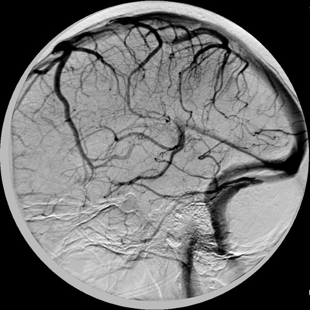
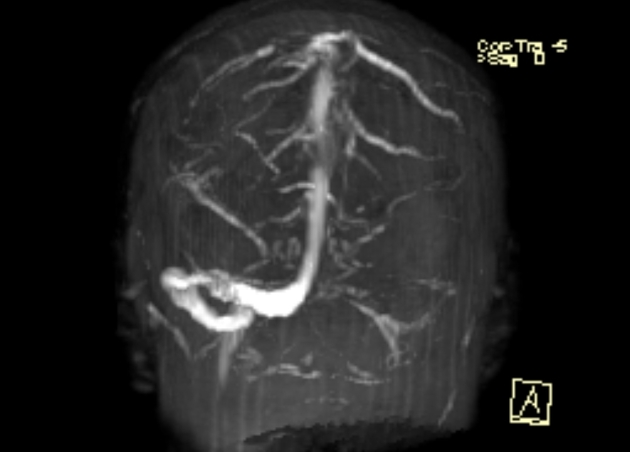
The superior anastomotic vein, also known as the vein of Trolard, is part of the superficial venous system of the brain.
It should not be confused with the venous circle of Trolard, the anastomotic venous circle of the base of the brain which is the inconsistently found venous homologue of the better-known arterial circle of Willis.
Gross anatomy
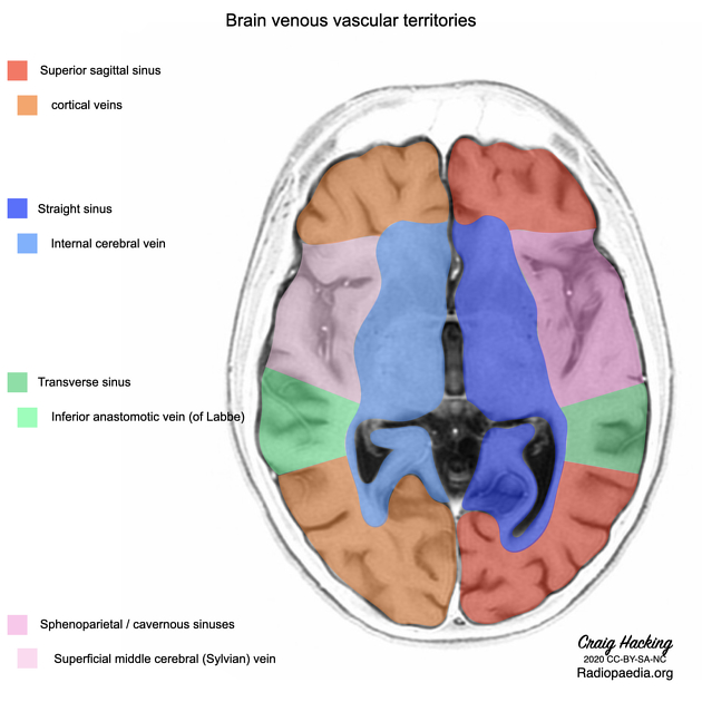
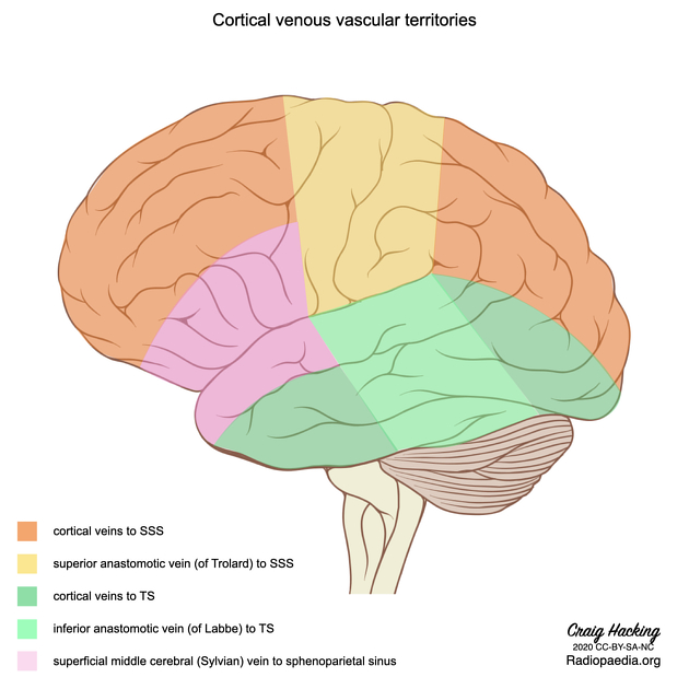
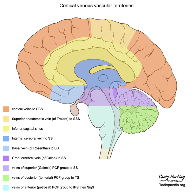
It is the largest superficial vein on the lateral surface of the parietal or frontal lobe that connects the superior sagittal sinus and the superficial middle cerebral vein (of Sylvius). It usually runs in the post-central sulcus 1 draining the adjacent cortex, but can be found in the central sulcus or anterior to it 5.
Its size is dictated by the relative size of the superficial middle cerebral vein and the inferior anastomotic vein of Labbé. The vein of Trolard is usually smaller than both of these. It has more variability than the vein of Labbé and is said to be the most variable of all the superficial veins of the brain 5.
As can be surmised by first principles, there is an inverse relationship between the size of the terminal Sylvian vein, the vein of Trolard, and the vein of Labbé, as all three share a similar drainage territory 3. Usually either the vein of Trolard or Labbé are dominant, both are small with the Sylvian vein being dominant or both are co-dominant with a small Sylvian vein. Occasionally the veins of Labbé and Trolard anastomose with themselves and bypass the Sylvain vein 5.
History and etymology
It is eponymously named after French surgeon and anatomist Jean Baptiste Paulin Trolard (1842–1910) who studied the anastomotic veins of the brain 3.
Trolard worked between France and Algeria and was an advocate of public vaccination in Algiers, establishing a vaccination institute alongside Louis Pasteur. He was also a strong critic of deforestation for which he created the League of Reforestation of Algeria 7.
Quiz questions
References
- 1. Standring S (editor). Gray's Anatomy (39th edition). Churchill Livingstone. (2011) ISBN:0443066841. Read it at Google Books - Find it at Amazon
- 2. Butler P, Mitchell A, Healy JC. Applied Radiological Anatomy. Cambridge University Press. (2012) ISBN:0521766664. Read it at Google Books - Find it at Amazon
- 3. Benner D, Hendricks B, Benet A, Lawton M. Eponyms in Vascular Neurosurgery: Comprehensive Review of 18 Veins. World Neurosurg. 2021;151:190-200. doi:10.1016/j.wneu.2021.05.053 - Pubmed
- 4. Marios Loukas, Misha Shea, Cory Shea, Martine Lutter-Hoppenheim, Paula Zand, R. Shane Tubbs, Aaron A. Cohen-Gadol. Jean Baptiste Paulin Trolard (1842–1910): his life and contributions to neuroanatomy: Historical vignette. (2010) Journal of Neurosurgery. 112 (6): 1192. doi:10.3171/2009.8.JNS09818
- 5. Tomasi S, Umana G, Scalia G et al. The Superficial Anastomosing Veins of the Human Brain Cortex: A Microneurosurgical Anatomical Study. Front Surg. 2022;8:817002. doi:10.3389/fsurg.2021.817002 - Pubmed
Incoming Links
- Venous haemorrhagic infarct due to cortical vein thrombosis
- Vein of Trolard rupture
- Disseminated medulloblastoma associated with dural venous sinus thrombosis
- Developmental venous anomaly
- Cerebral venous thrombosis
- Venous infarct due to superior sagittal sinus and superior cortical vein thrombosis
- Extradural spinal CSF leak
- Cortical vein thrombosis with infarct and haemorrhage
- Vein of Trolard thrombosis with venous infarction (CT perfusion)
- Venous vascular territories of the lateral cerebral cortex (illustration)
- Brain venous vascular territories (diagram)
- Cerebral arteriovenous malformation
- Polymicrogyria
- Cerebral venous infarct
- Veins of Trolard and Rosenthal
- Cerebral arteriovenous malformation
- Veins of Labbé and Trolard
Related articles: Anatomy: Brain
-
brain
- grey matter
- white matter
-
cerebrum
-
cerebral hemisphere (telencephalon)
- cerebral lobes and gyri
- frontal lobe
- parietal lobe
-
occipital lobe
- occipital pole
- lingual gyrus
- fusiform gyrus (Brodmann area 37)
- calcarine (visual) cortex
- cuneus
- temporal lobe
- basal forebrain
- limbic system
- insula
-
cerebral sulci and fissures (A-Z)
- calcarine fissure
- callosal sulcus
- central (Rolandic) sulcus
- cingulate sulcus
- collateral sulcus
- inferior frontal sulcus
- inferior occipital sulcus
- inferior temporal sulcus
- interhemispheric fissure
- intraparietal sulcus
- lateral (Sylvian) sulcus
- lateral occipital sulcus
- marginal sulcus
- occipitotemporal sulcus
- olfactory sulcus
- paracentral sulcus
- paraolfactory sulcus
- parieto-occipital fissure
- posterior parolfactory sulcus
- precentral sulcus
- preoccipital notch
- postcentral sulcus
- rhinal sulcus
- rostral sulcus
- subparietal sulcus
- superior frontal sulcus
- superior occipital sulcus
- superior temporal sulcus
- cortical histology
- cerebral lobes and gyri
- white matter tracts
- deep grey matter
-
pituitary gland
- posterior pituitary and stalk (part of diencephalon)
- anterior pituitary
- inferior hypophyseal arterial circle
- diencephalon
-
cerebral hemisphere (telencephalon)
-
brainstem
- midbrain (mesencephalon)
- pons (part of metencephalon)
- medulla oblongata (myelencephalon)
- white matter
-
grey matter
- non-cranial nerve
-
cranial nerve nuclei
- oculomotor nucleus
- Edinger-Westphal nucleus
- trochlear nucleus
- motor nucleus of CN V
- mesencephalic nucleus of CN V
- main sensory nucleus of CN V
- spinal nucleus of CN V
- abducent nucleus
- facial nucleus
- superior salivatory nucleus
- cochlear nuclei
- vestibular nuclei
- inferior salivatory nucleus
- solitary tract nucleus
- ambiguus nucleus
- dorsal vagal motor nucleus
- hypoglossal nucleus
-
cerebellum (part of metencephalon)
- vermis
- cerebellar hemisphere
- cerebellar peduncles
- cranial meninges (meninx primitiva)
- CSF spaces
-
cranial nerves (mnemonic)
- olfactory nerve (CN I)
- optic nerve (CN II)
- oculomotor nerve (CN III)
- trochlear nerve (CN IV)
- trigeminal nerve (CN V) (mnemonic)
- abducens nerve (CN VI)
- facial nerve (CN VII) (segments mnemonic | branches mnemonic)
-
vestibulocochlear nerve (CN VIII)
- vestibular ganglion (Scarpa's ganglion)
- glossopharyngeal nerve (CN IX)
- vagus nerve (CN X)
- spinal accessory nerve (CN XI)
- hypoglossal nerve (CN XII)
- functional neuroanatomy
- CNS development
- cerebral vascular supply
- arteries
- vascular territories
-
circle of Willis
- internal carotid artery (ICA) (segments)
- vertebral artery
-
normal variants
- intracranial arterial fenestration
- internal carotid artery (ICA)
- anterior cerebral artery (ACA)
- middle cerebral artery (MCA)
- posterior cerebral artery (PCA)
- basilar artery
- persistent carotid-vertebrobasilar artery anastomoses (mnemonic)
- vertebral artery
- ophthalmic artery
-
cerebral venous system
-
dural venous sinuses
- basilar venous plexus
- cavernous sinus (mnemonic)
- clival diploic veins
- inferior petro-occipital vein
- inferior petrosal sinus
- inferior sagittal sinus
- intercavernous sinus
- internal carotid artery venous plexus of Rektorzik
- jugular bulb
- marginal sinus
- occipital sinus
- sigmoid sinus
- sphenoparietal sinus
- straight sinus
- superior petrosal sinus
- superior sagittal sinus
- torcula herophili
- transverse sinus
-
cerebral veins
-
superficial veins of the brain
- superior cerebral veins (superficial cerebral veins)
- inferior cerebral veins
- superficial middle cerebral vein
- superior anastomotic vein (of Trolard)
- inferior anastomotic vein (of Labbe)
-
superficial veins of the brain
-
deep veins of the brain
- great cerebral vein (of Galen)
- venous circle of Trolard
- normal variants
-
dural venous sinuses
- arteries
- glymphatic pathway


 Unable to process the form. Check for errors and try again.