
Calcaneal fracture
Citation, DOI, disclosures and article data
At the time the article was created The Radswiki had no recorded disclosures.
View The Radswiki's current disclosuresAt the time the article was last revised Gabriel Orlando González Herrera had no financial relationships to ineligible companies to disclose.
View Gabriel Orlando González Herrera's current disclosures- Calcaneal fractures
- Fractures of the calcaneus
- Fracture involving the calcaneus
- Fractured calcaneus
- Fracture of calcaneus
Calcaneal fractures are the most common tarsal fracture and can occur in a variety of settings.
On this page:
Epidemiology
The calcaneus is the most commonly fractured tarsal bone and accounts for about 2% of all fractures 2 and ~60% of all tarsal fractures 3.
Pathology
Calcaneal fractures can be divided broadly into two types depending on whether there is articular involvement of the subtalar joint 2,7,8:
-
extra-articular: 25-30%
-
extra-articular body fracture
medial sustentaculum
-
intra-articular: 70-75%
intra-articular body fracture
The calcaneus is also a common site of stress fractures, occurring in the posterosuperior aspect.
Another method of classification is as
type A fractures: the anterior process of the calcaneus is fractured
type B: fracture of the mid calcaneus, trochlear process, and sustentaculum tali
type C: fracture of the posterior tuberosity
Radiographic features
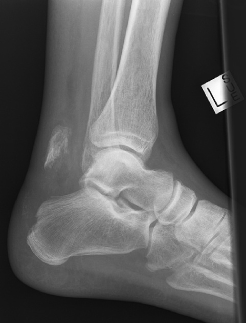
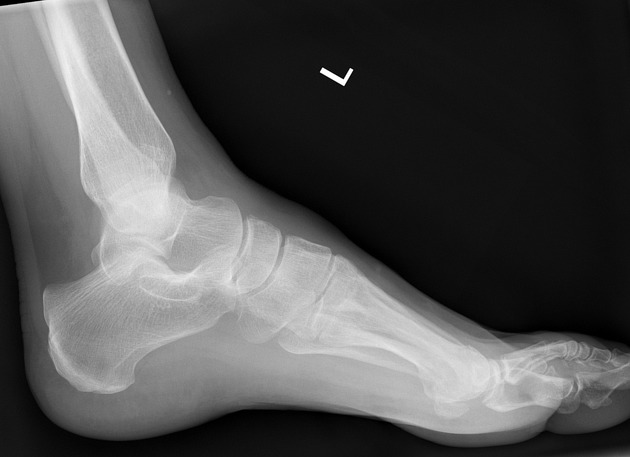
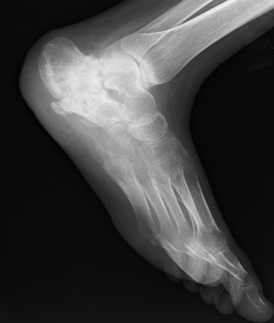
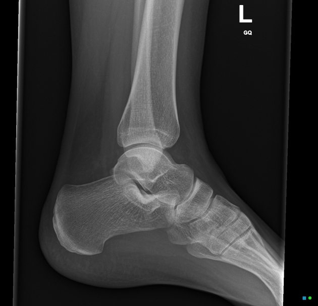
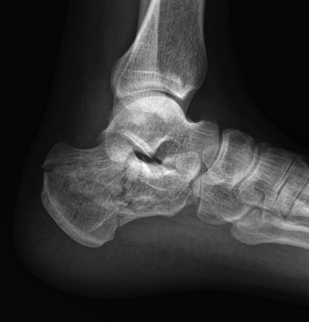
Plain radiograph
Calcaneal fractures are best assessed with a calcaneal series of radiographs, though are often identified on a lateral ankle radiograph if the presentation does not lead the requesting of calcaneal views specifically. The Böhler and Gissane angles are used to assess the severity of calcaneal fractures, and their postoperative appearance is correlated with functional outcomes 12.
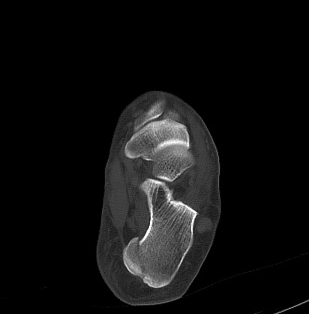
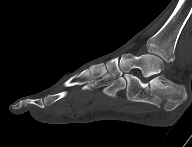
CT
CT is the modality of choice to evaluate calcaneal fracture. It can show the extent and extra- or intra-articular components of the fracture and hematoma along the sole of the foot (Mondor sign). Intra-articular fractures are often classified using the Sanders classification system, which is one of the only systems that correlates well with patient outcome.
Practical points
If bilateral calcaneal fractures are seen, then the spine should also be evaluated for fracture as the mechanism of injury is often a large load to the axial skeleton, such as a fall from height.
If an intra-articular calcaneal fracture is seen, the images should be scrutinized for a lateral malleolar fleck sign (ankle), which raises the likelihood of peroneal tendon instability 10.
Quiz questions

References
- 1. Borut Marincek, Robert F. Dondelinger. Emergency Radiology. (2006) ISBN: 9783540262275 - Google Books
- 2. Daftary A, Haims A, Baumgaertner M. Fractures of the Calcaneus: A Review with Emphasis on CT. Radiographics. 2005;25(5):1215-26. doi:10.1148/rg.255045713 - Pubmed
- 3. Badillo K, Pacheco J, Padua S, Gomez A, Colon E, Vidal J. Multidetector CT Evaluation of Calcaneal Fractures. Radiographics. 2011;31(1):81-92. doi:10.1148/rg.311105036 - Pubmed
- 4. Heger L, Wulff K, Seddiqi M. Computed Tomography of Calcaneal Fractures. AJR Am J Roentgenol. 1985;145(1):131-7. doi:10.2214/ajr.145.1.131 - Pubmed
- 5. Matherne T, Tivorsak T, Monu J. Calcaneal Fractures: What the Surgeon Needs to Know. Curr Probl Diagn Radiol. 2007;36(1):1-10. doi:10.1067/j.cpradiol.2006.07.006 - Pubmed
- 6. Wechsler R, Schweitzer M, Karasick D, Deely D, Morrison W. Helical CT of Calcaneal Fractures: Technique and Imaging Features. Skeletal Radiol. 1998;27(1):1-6. doi:10.1007/s002560050325 - Pubmed
- 7. Bruce D. Browner. Skeletal Trauma. (2009) ISBN: 9781416022206 - Google Books
- 8. Jacob Mandell. Core Radiology. (2013) ISBN: 9781107679689 - Google Books
- 9. Lee A. Grant, Nyree Griffin. Grainger & Allison's Diagnostic Radiology Essentials E-Book. (2018) ISBN: 9780323568845 - Google Books
- 10. Mahmoud K, Mekhaimar M, Alhammoud A. Prevalence of Peroneal Tendon Instability in Calcaneus Fractures: A Systematic Review and Meta-Analysis. J Foot Ankle Surg. 2018;57(3):572-8. doi:10.1053/j.jfas.2017.11.032 - Pubmed
- 11. Yu S & Yu J. Calcaneal Avulsion Fractures: An Often Forgotten Diagnosis. AJR Am J Roentgenol. 2015;205(5):1061-7. doi:10.2214/AJR.14.14190 - Pubmed
- 12. Su Y, Chen W, Zhang T, Wu X, Wu Z, Zhang Y. Bohler's Angle's Role in Assessing the Injury Severity and Functional Outcome of Internal Fixation for Displaced Intra-Articular Calcaneal Fractures: A Retrospective Study. BMC Surg. 2013;13(1):40. doi:10.1186/1471-2482-13-40 - Pubmed
Incoming Links
- Retrocalcaneal bursitis
- Tarsal tunnel syndrome
- Trauma
- CT ankle (protocol)
- Böhler angle
- Subtalar arthritis
- Calcaneus
- Sural neuropathy
- Sanders CT classification of calcaneal fracture
- Musculoskeletal curriculum
- Lumbar spine fracture
- Ankle radiograph (checklist)
- Ankle radiograph (an approach)
- Tarsal fracture
- Fleck sign (ankle)
- Lover's fracture
- Gissane angle
- Calcaneal tuberosity avulsion fracture
- Extra-articular lateral hindfoot impingement syndrome
- Mondor sign (foot)
- Calcaneal fracture
- Extra-articular calcaneal fracture
- Lover’s fracture
- Calcaneal stress fracture
- Calcaneus fracture
- Sever disease (calcaneal apophysitis)
- Stress fractures of both calcanei
- Bilateral calcaneal fractures
- Calcaneal fracture
- Beak type calcaneus fracture
- Calcaneal fracture
- Calcaneal fracture - tongue-type
- Calcaneal fracture - Sanders type 3ab
- Os calcaneus secundarius
- Böhler angle
- Neglected congenital talipes equinovarus
- Calcaneus fracture
- Calcaneal fracture
- Calcaneal fracture - Sanders type 4
- Calcaneus fracture
Related articles: Fractures
-
fracture
- terminology[+][+]
- fracture location
- diaphyseal fracture
- metaphyseal fracture
- physeal fracture
- epiphyseal fracture
- fracture types
- avulsion fracture
- articular surface injuries
- complete fracture
- incomplete fracture
- infraction
- compound fracture
- pathological fracture
- stress fracture
- fracture displacement
- fracture location
- fracture healing[+][+]
- skull fractures[+][+]
-
facial fractures[+][+]
- fractures involving a single facial buttress
- alveolar process fractures
- frontal sinus fracture
- isolated zygomatic arch fractures
- mandibular fracture
- nasal bone fracture
- orbital blow-out fracture
- paranasal sinus fractures
- complex fractures
- dental fractures
- fractures involving a single facial buttress
-
spinal fractures[+][+]
- classification (AO Spine classification systems)
-
cervical spine fracture classification systems
- AO classification of upper cervical injuries
- AO classification of subaxial injuries
- Anderson and D'Alonzo classification (odontoid fracture)
- Roy-Camille classification (odontoid process fracture)
- Gehweiler classifcation (atlas fractures)
- Levine and Edwards classification (hangman fracture)
- Allen and Ferguson classification (subaxial spine injuries)
- subaxial cervical spine injury classification (SLIC)
- thoracolumbar spinal fracture classification systems
- three column concept of spinal fractures (Denis classification)
- classification of sacral fractures
-
cervical spine fracture classification systems
- spinal fractures by region
- spinal fracture types
- classification (AO Spine classification systems)
- rib fractures[+][+]
- sternal fractures
-
upper limb fractures[+][+]
- classification
- Rockwood classification (acromioclavicular joint injury)
- AO classification (clavicle fracture)
- Neer classification (clavicle fracture)
- Neer classification (proximal humeral fracture)
- AO classification (proximal humeral fracture)
- AO/OTA classification of distal humeral fractures
- Milch classification (lateral humeral condyle fracture)
- Weiss classification (lateral humeral condyle fracture)
- Bado classification of Monteggia fracture-dislocations (radius-ulna)
- Mason classification (radial head fracture)
- Frykman classification (distal radial fracture)
- Mayo classification (scaphoid fracture)
- Hintermann classification (gamekeeper's thumb)
- Eaton classification (volar plate avulsion injury)
- Keifhaber-Stern classification (volar plate avulsion injury)
- upper limb fractures by region
- shoulder
- clavicular fracture
-
scapular fracture
- acromion fracture
- coracoid process fracture
- glenoid fracture
- humeral head fracture
- proximal humeral fracture
- humeral neck fracture
- arm
- elbow
- forearm
- wrist
-
carpal bones
- scaphoid fracture
- lunate fracture
- capitate fracture
- triquetral fracture
- pisiform fracture
- hamate fracture
- trapezoid fracture
- trapezium fracture
- hand
- shoulder
- classification
- lower limb fractures
- classification by region[+][+]
- pelvic fractures
- hip fractures
- Pipkin classification (femoral head fracture)
- Garden classification (hip fracture)
- American Academy of Orthopedic Surgeons classification (periprosthetic hip fracture)
- Cooke and Newman classification (periprosthetic hip fracture)
- Johansson classification (periprosthetic hip fracture)
- Vancouver classification (periprosthetic hip fracture)
- femoral
- knee
- Schatzker classification (tibial plateau fracture)
- AO classification of distal femur fractures
- Meyers and McKeevers classification (anterior cruciate ligament avulsion fracture)
- tibia/fibula
- Watson-Jones classification (tibial tuberosity avulsion fracture)
- ankle
- foot
- Berndt and Harty classification (osteochondral lesions of the talus)
- Sanders CT classification (calcaneal fracture)
- Hawkins classification (talar neck fracture)
- Myerson classification (Lisfranc injury)
- Nunley-Vertullo classification (Lisfranc injury)
- pelvis and lower limb fractures by region
- pelvic fracture[+][+]
- sacral fracture[+][+]
- coccygeal fracture
-
hip[+][+]
- acetabular fracture
- femoral head fracture
-
femoral neck fracture
- subcapital fracture
- transcervical fracture
- basicervical fracture
-
trochanteric fracture
- pertrochanteric fracture
- intertrochanteric fracture
- subtrochanteric fracture
- femur[+][+]
- mid-shaft fracture
- bisphosphonate-related fracture
- distal femoral fracture
- knee[+][+]
- avulsion fractures
- Segond fracture
- reverse Segond fracture
- anterior cruciate ligament avulsion fracture
- posterior cruciate ligament avulsion fracture
- arcuate complex avulsion fracture (arcuate sign)
- biceps femoris avulsion fracture
- iliotibial band avulsion fracture
- semimembranosus tendon avulsion fracture
- Stieda fracture (MCL avulsion fracture)
- patellar fracture
- tibial plateau fracture
- avulsion fractures
- leg[+][+]
- tibial tuberosity avulsion fracture
- tibial shaft fracture
- fibular shaft fracture
- Maisonneuve fracture
- ankle[+][+]
- foot
- tarsal bones
- metatarsal bones[+][+]
- phalanges
- classification by region[+][+]
- terminology[+][+]


 Unable to process the form. Check for errors and try again.