
Extraconal orbital compartment
Citation, DOI, disclosures and article data
At the time the article was created Craig Hacking had no recorded disclosures.
View Craig Hacking's current disclosuresAt the time the article was last revised Craig Hacking had the following disclosures:
- Philips Australia, Paid speaker at Philips Spectral CT events (ongoing)
These were assessed during peer review and were determined to not be relevant to the changes that were made.
View Craig Hacking's current disclosures- Extraconal space
- Extraconal orbital space
- Extraconal compartment
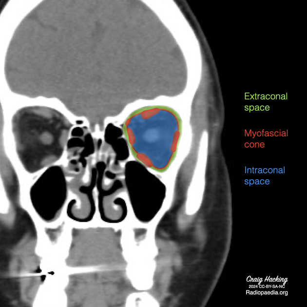
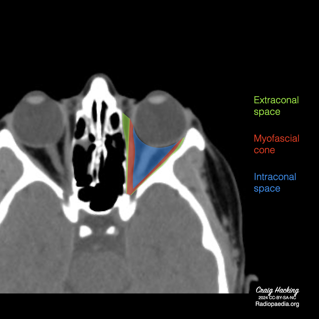
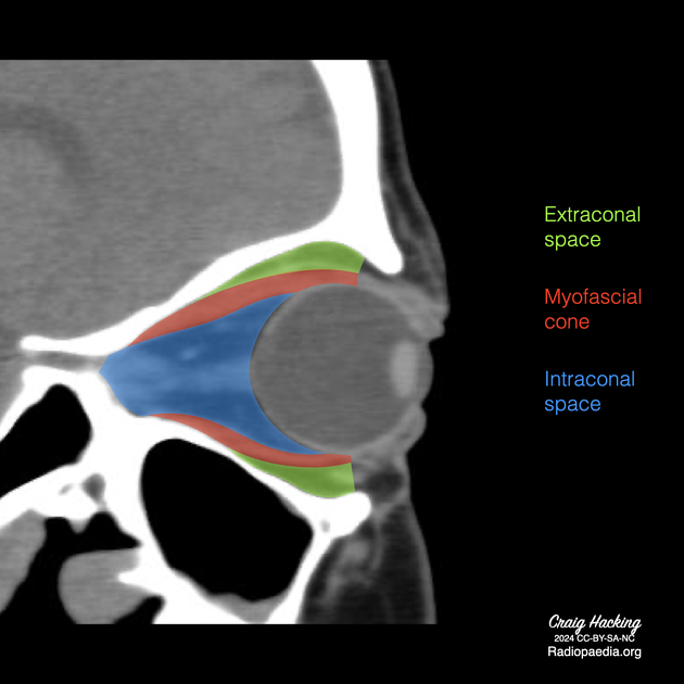
The extraconal orbital compartment or extraconal space is the space within the orbit outside the musculofascial cone. The base of which is anterior and is formed by the orbital septum that surrounds the equator of the globe. The external sides are formed by the bones of the orbit and their periosteum. The internal sides are formed by the extraocular muscles and their surrounding fascia which pass posteriorly and converge on the tendinous ring at the orbital apex. Internal to this space is the intraconal space.
Contents
-
nerves
trochlear nerve (CN IV)
-
branches of the ophthalmic division of the trigeminal nerve
-
nasociliary nerve is intraconal, but gives extraconal branches:
-
branches of the maxillary division of the trigeminal nerve
infra-orbital nerve (before entering the infra-orbital canal)
-
arteries
-
extraconal branches of the ophthalmic artery that have an intraconal origin:
infra-orbital artery (before entering the infra-orbital canal)
-
-
veins
infra-orbital vein (before entering the infra-orbital canal)
fat
ADVERTISEMENT: Supporters see fewer/no ads
Related pathology
References
- 1. Moore KL, Agur AMR, Dalley AF. Clinically oriented anatomy. LWW. ISBN:1451119453. Read it at Google Books - Find it at Amazon
- 2. Last's anatomy, regional and applied. Churchill Livingstone. ISBN:044304662X. Read it at Google Books - Find it at Amazon
- 3. Butler P, Mitchell A, Healy JC. Applied Radiological Anatomy. Cambridge University Press. (2012) ISBN:0521766664. Read it at Google Books - Find it at Amazon
Incoming Links
- Anterior ethmoidal nerve
- Orbital infection
- Orbital spaces
- Orbital metastasis
- Orbit
- Orbital dermoid cyst
- Zygomaticotemporal nerve
- Extraconal orbital lesions
- Posterior ethmoidal nerve
- Zygomaticofacial nerve
- Orbital lymphoproliferative lesions
- Zygomatic nerve
- Nasociliary nerve
- Myofascial cone
- Infraorbital nerve
- Inferior ophthalmic vein
- Ocular globe
- Supraorbital nerve
- Infratrochlear nerve
- Lacrimal gland
- Orbital spaces (CT)
- Intraorbital wood foreign body
- Orbital metastasis of breast cancer
- Frontoethmoidal sinus osteoma
- Cystic lymphangioma of the orbital cavity
- Fronto-ethmoidal sinus mucocele
- Idiopathic orbital inflammation
- Subperiosteal abscess of the orbit
- Orbital blow-out fracture with extraconal emphysema and proptosis
- Ocular foreign body
- En plaque meningioma with associated CPA / frontal convexity meningiomas
- Adenoid cystic carcinoma - maxillary sinus
Related articles: Anatomy: Head and neck
- skeleton of the head and neck
-
cranial vault
- scalp (mnemonic)
- fontanelle
-
sutures
- calvarial
- facial
- frontozygomatic suture
- frontomaxillary suture
- frontolacrimal suture
- frontonasal suture
- temporozygomatic suture
- zygomaticomaxillary suture
- parietotemporal suture (parietomastoid suture)
- occipitotemporal suture (occipitomastoid suture)
- sphenofrontal suture
- sphenozygomatic suture
- spheno-occipital suture (not a true suture)
- lacrimomaxillary suture
- nasomaxillary suture
- internasal suture
- basal/internal
- skull landmarks
- frontal bone
- temporal bone
- parietal bone
- occipital bone
- skull base (foramina)
-
facial bones
- midline single bones
- paired bilateral bones
- cervical spine
- hyoid bone
- laryngeal cartilages
-
cranial vault
- muscles of the head and neck
- muscles of the tongue (mnemonic)
- muscles of mastication
-
facial muscles
- epicranius muscle
- circumorbital and palpebral muscles
- nasal muscles
-
buccolabial muscles
- elevators, retractors and evertors of the upper lip
- levator labii superioris alaeque nasalis muscle
- levator labii superioris muscle
- zygomaticus major muscle
- zygomaticus minor muscle
- levator anguli oris muscle
- malaris muscle
- risorius muscle
- depressors, retractors and evertors of the lower lip
- depressor labii inferioris muscle
- depressor anguli oris muscle
- mentalis muscle
- compound sphincter
-
orbicularis oris muscle
- incisivus labii superioris muscle
- incisivus labii inferioris muscle
-
orbicularis oris muscle
- muscle of mastication
- modiolus
- elevators, retractors and evertors of the upper lip
- muscles of the middle ear
- orbital muscles
- muscles of the soft palate
- pharyngeal muscles
- suprahyoid muscles
- infrahyoid muscles
- intrinsic muscles of the larynx
- muscles of the neck
- platysma muscle
- longus colli muscle
- longus capitis muscle
- scalenus anterior muscle
- scalenus medius muscle
- scalenus posterior muscle
- scalenus pleuralis muscle
- sternocleidomastoid muscle
-
suboccipital muscles
- rectus capitis posterior major muscle
- rectus capitis posterior minor muscle
- obliquus capitis superior muscle
- obliquus capitis inferior muscle
- accessory muscles of the neck
- deep cervical fascia
-
deep spaces of the neck
- anterior cervical space
- buccal space
- carotid space
- danger space
- deep cervical fascia
- infratemporal fossa
- masticator space
- parapharyngeal space
- stylomandibular tunnel
- parotid space
- pharyngeal (superficial) mucosal space
- perivertebral space
- posterior cervical space
- pterygopalatine fossa
- retropharyngeal space
- suprasternal space (of Burns)
- visceral space
- surgical triangles of the neck
- orbit
- ear
- paranasal sinuses
- upper respiratory tract
- viscera of the neck
- blood supply of the head and neck
-
arterial supply
-
common carotid artery
- carotid body
- carotid bifurcation
- subclavian artery
- variants
-
common carotid artery
- venous drainage
-
arterial supply
- innervation of the head and neck
-
cranial nerves
- olfactory nerve (CN I)
- optic nerve (CN II)
- oculomotor nerve (CN III)
- trochlear nerve (CN IV)
-
trigeminal nerve (CN V) (mnemonic)
- trigeminal ganglion
- ophthalmic division
- maxillary division
- mandibular division
- abducens nerve (CN VI)
- facial nerve (CN VII)
-
vestibulocochlear nerve (CN VIII)
- vestibular ganglion (Scarpa's ganglion)
- glossopharyngeal nerve (CN IX)
- vagus nerve (CN X)
- (spinal) accessory nerve (CN XI)
- hypoglossal nerve (CN XII)
- parasympathetic ganglia of the head and neck
- cervical sympathetic ganglia
- greater occipital nerve
- third occipital nerve
-
cervical plexus
- muscular branches
- longus capitis
- longus colli
- scalenes
- geniohyoid
- thyrohyoid
-
ansa cervicalis
- omohyoid (superior and inferior bellies separately)
- sternothyroid
- sternohyoid
- phrenic nerve
- contribution to the accessory nerve (CN XI)
- cutaneous branches
- muscular branches
- brachial plexus
- pharyngeal plexus
-
cranial nerves
- lymphatic drainage of the head and neck
- embryological development of the head and neck


 Unable to process the form. Check for errors and try again.