
Rotator cuff interval
Citation, DOI, disclosures and article data
Citation:
Gaillard F, Elfeky M, Jabaz D, et al. Rotator cuff interval. Reference article, Radiopaedia.org (Accessed on 31 Mar 2025) https://doi.org/10.53347/rID-1996
Permalink:
rID:
1996
Article created:
Disclosures:
At the time the article was created Frank Gaillard had no recorded disclosures.
View Frank Gaillard's current disclosures
Last revised:
Disclosures:
At the time the article was last revised Mostafa Elfeky had no financial relationships to ineligible companies to disclose.
View Mostafa Elfeky's current disclosures
Revisions:
13 times, by
8 contributors -
see full revision history and disclosures
Systems:
Tags:
Synonyms:
- Rotator interval
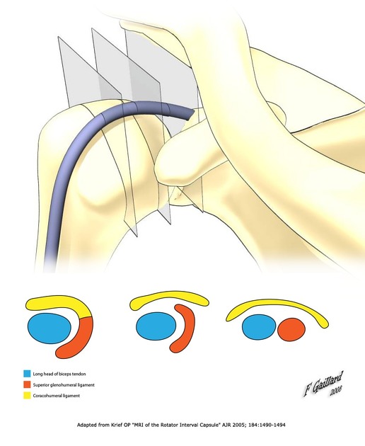
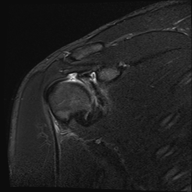
The rotator cuff interval is a triangular space between the tendons of subscapularis and supraspinatus and the base of the coracoid process.
On this page:
Gross anatomy
The combination of the coracohumeral ligament and superior glenohumeral ligament has a complex relationship with the long head of biceps tendon (LHB), acting together to prevent subluxation or anterior dislocation of the LHB.
Boundaries
- anterior: posterior aspect of the subscapularis tendon
- posterior: anterior border of the supraspinatus tendon
- medial: lateral margin of the base of the coracoid process
- roof: the rotator interval capsule, reinforced by:
- coracohumeral ligament
Contents
- tendon of the long head of biceps brachii
- superior glenohumeral ligament
History and etymology
It was first defined by C S Neer in 1970 3.
Related pathology
- rotator interval tear
- long head of biceps tendon dislocation
- rotator interval changes in adhesive capsulitis
Quiz questions
References
- 1. Krief O. MRI of the Rotator Interval Capsule. AJR Am J Roentgenol. 2005;184(5):1490-4. doi:10.2214/ajr.184.5.01841490 - Pubmed
- 2. Petchprapa C, Beltran L, Jazrawi L, Kwon Y, Babb J, Recht M. The Rotator Interval: A Review of Anatomy, Function, and Normal and Abnormal MRI Appearance. AJR Am J Roentgenol. 2010;195(3):567-76. doi:10.2214/AJR.10.4406 - Pubmed
- 3. Frank R, Taylor D, Verma N, Romeo A, Mologne T, Provencher M. The Rotator Interval of the Shoulder: Implications in the Treatment of Shoulder Instability. Orthop J Sports Med. 2015;3(12):2325967115621494. doi:10.1177/2325967115621494 - Pubmed
- 4. Tamborrini G, Möller I, Bong D et al. The Rotator Interval - A Link Between Anatomy and Ultrasound. Ultrasound Int Open. 2017;3(3):E107-16. doi:10.1055/s-0043-110473 - Pubmed
Incoming Links
Articles:
- Biceps brachii muscle
- Coracoacromial arch
- Subscapularis tendon tear
- Biceps pulley injury
- Shoulder protocol (MRI)
- Massive rotator cuff tear
- Glenohumeral joint injection (technique)
- Adhesive capsulitis of the shoulder
- Ultrasound of the shoulder
- Long head of biceps tendon dislocation
- Subacromial-subdeltoid bursa
- Biceps pulley
- Glenoid labrum
- Long head of biceps tendon
- Supraspinatus tendon tear
- MRI of the shoulder (an approach)
- Coracohumeral ligament
- Rotator cuff
- Rotator cuff tear
Cases:
Multiple choice questions:
Related articles: Anatomy: Upper limb
-
skeleton of the upper limb
- clavicle
- scapula
- humerus
- radius
- ulna
- hand
- accessory ossicles of the upper limb
- accessory ossicles of the shoulder
- accessory ossicles of the elbow
-
accessory ossicles of the wrist (mnemonic)
- os centrale carpi
- os epilunate
- os epitriquetrum
- os styloideum
- os hamuli proprium
- lunula
- os triangulare
- trapezium secondarium
- os paratrapezium
- os radiostyloideum (persistent radial styloid)
- joints of the upper limb
-
pectoral girdle
-
shoulder joint
- articulations
- associated structures
- joint capsule
- bursae
- ligaments
- movements
- scapulothoracic joint
-
glenohumeral joint
- arm flexion
- arm extension
- arm abduction
- arm adduction
- arm internal rotation (medial rotation)
- arm external rotation (lateral rotation)
- circumduction
- arterial supply - scapular anastomosis
- ossification centers
-
shoulder joint
-
elbow joint
- proximal radioulnar joint
- ligaments
- associated structures
- movements
- alignment
- arterial supply - elbow anastomosis
- development
-
wrist joint
- articulations
-
ligaments
- intrinsic ligaments
- extrinsic ligaments
- radioscaphoid ligament
- dorsal intercarpal ligament
- dorsal radiotriquetral ligament
- dorsal radioulnar ligament
- volar radioulnar ligament
- radioscaphocapitate ligament
- long radiolunate ligament
- Vickers ligament
- short radiolunate ligament
- ulnolunate ligament
- ulnotriquetral ligament
- ulnocapitate ligament
- ulnar collateral ligament
- associated structures
- extensor retinaculum
- flexor retinaculum
- joint capsule
- movements
- alignment
- ossification centers
-
hand joints
- articulations
- carpometacarpal joint
-
metacarpophalangeal joints
- palmar ligament (plate)
- collateral ligament
-
interphalangeal joints
- palmar ligament (plate)
- collateral ligament
- movements
- ossification centers
- articulations
-
pectoral girdle
- spaces of the upper limb
- muscles of the upper limb
- shoulder girdle
- anterior compartment of the arm
- posterior compartment of the arm
-
anterior compartment of the forearm
- superficial
- intermediate
- deep
-
posterior compartment of the forearm (extensors)
- superficial
- deep
- muscles of the hand
-
accessory muscles
- elbow
- volar wrist midline
- palmaris longus profundus
- aberrant palmaris longus
- volar wrist radial-side
- accessory flexor digitorum superficialis indicis
- flexor indicis profundus
- flexor carpi radialis vel profundus
- accessory head of the flexor pollicis longus (Gantzer muscle, common)
- volar wrist ulnar-side
- dorsal wrist
- blood supply to the upper limb
-
arteries
- subclavian artery (mnemonic)
- axillary artery
- brachial artery (proximal portion)
- ulnar artery
- radial artery
- veins
-
arteries
- innervation of the upper limb
- intercostobrachial nerve
-
brachial plexus (mnemonic)
- branches from the roots
- branches from the trunks
- branches from the cords
- lateral cord
- posterior cord
- medial cord
- terminal branches
- lymphatic drainage of the upper limb


 Unable to process the form. Check for errors and try again.