
The internal capsule (TA: capsula interna) is a deep subcortical structure that contains a concentration of afferent and efferent white matter projection fibers. Anatomically, this is an important area because of the high concentration of both motor and sensory projection fibers 1,2. Afferent fibers pass from cell bodies of the thalamus to the cortex, and efferent fibers pass from cell bodies of the cortex to the cerebral peduncle of the midbrain 2. Fibers from the internal capsule contribute to the corona radiata.
On this page:
Gross anatomy
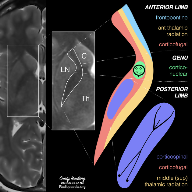
The internal capsule is made up of five parts. These are the anterior limb, genu, posterior limb, retrolentiform and sublentiform parts of the internal capsule 1,2:
-
anterior limb (anterior crus)
lies between the head of the caudate nucleus medially and the lentiform nucleus laterally
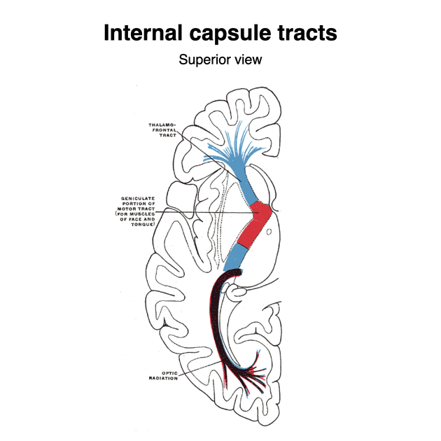
contains the anterior thalamic radiation and frontopontine fibers
-
genu
lies medial to the apex of the lentiform nucleus
contains corticonuclear fibers (previously called corticobulbar fibers)
-
posterior limb (posterior crus)
lies between the thalamus medially and the lentiform nucleus laterally
-
contains the
-
corticospinal fibers lying in the anterior two-thirds of the posterior limb
fibers from anterior to posterior: head, arm, hand, trunk, leg, perineum 2
middle (superior) thalamic radiation which contains somatosensory fibers from the ventral posterior thalamic nucleus
-
-
retrolentiform part
lies posterior to the lentiform nucleus
-
contains the
geniculocalcarine or optic radiation (posterior thalamic radiation) from the lateral geniculate nucleus
corticopontine fibers (parietopontine and occiptopontine fibers) 2
-
sublentiform part
lies inferior to the lentiform nucleus
contains the auditory radiations (inferior thalamic radiation) from the medial geniculate nucleus)and temporopontine fibers
Arterial supply
The blood supply of the internal capsule is variable but is commonly from small perforating branches of the middle cerebral artery and anterior cerebral artery. These include the lateral lenticulostriate arteries and the recurrent artery of Heubner respectively 3. In addition, the anterior choroidal artery from the internal carotid artery supplies the posterior limb and retrolentiform part of the internal capsule 3,4.
Radiographic features
CT
best appreciated on axial images at the level of the insular cortex
appears relatively hypodense to surrounding basal ganglia structures
MRI
in term neonates, internal capsule appears as higher T1-weighted and lower T2-weighted intensity when compared to basal ganglia and thalamus 6


 Unable to process the form. Check for errors and try again.