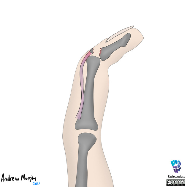
Mallet finger
Mallet finger refers to injuries of the extensor mechanism of the finger at the level of the distal interphalangeal joint (DIP). They are the most prevalent finger tendon injury in sport. They may represent an isolated tendinous injury or occur in combination with an avulsion fracture of the dorsal base of the distal phalanx.
On this page:
Clinical presentation
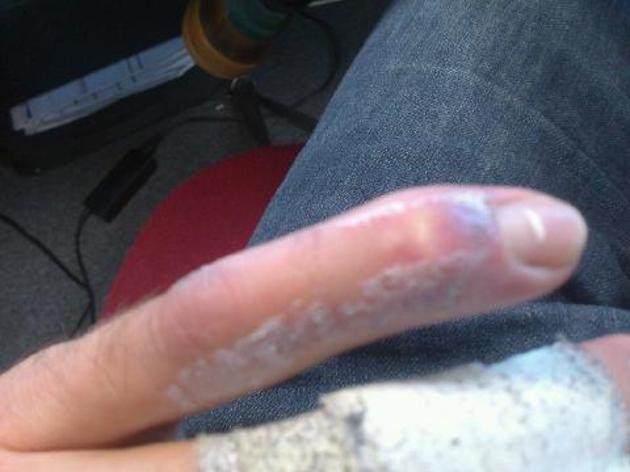
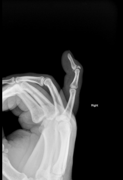
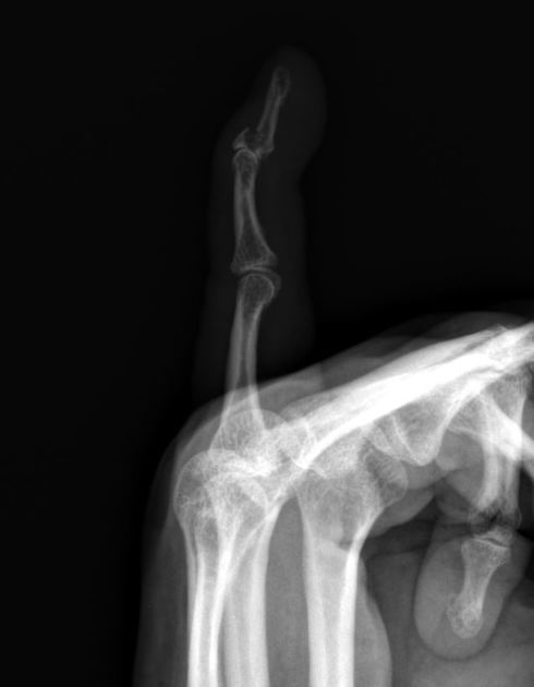
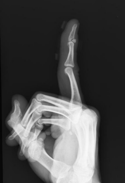
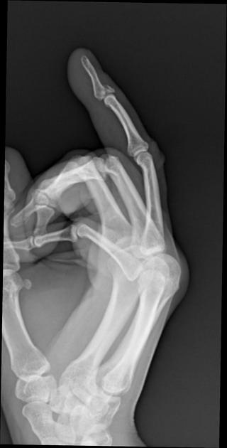
It is characterized by an inability to extend the finger at the distal interphalangeal (DIP) joint. There is slight flexion at this joint, which is where the term "mallet" comes from - the finger position resembles a mallet (for example, a piano key mallet).
The injury classically occurs while playing sports where the DIP undergoes sudden flexion (extended finger is struck at the tip by an object, e.g. baseball, basketball), or a crush injury (slamming a door towards the distal interphalangeal joint) in the extensor direction.
Pathology
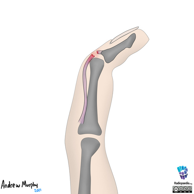
The DIP joint is extended by combined pulling force of the terminal (lateral bands) of the extensor tendon, functioning together with the oblique retinacular ligament 7. Injury to these structures commonly results from direct axial or flexion loading of the DIP joint, as can occur by direct blow from a ball.
The terminal extensor tendon inserts on the DIP joint capsule, and so injurious force may also result in intra-articular avulsion fracture of the base of the distal phalanx. This may represent an epiphyseal injury in skeletally-immature children 7.
Radiographic features
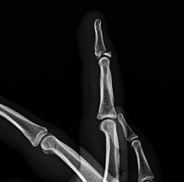
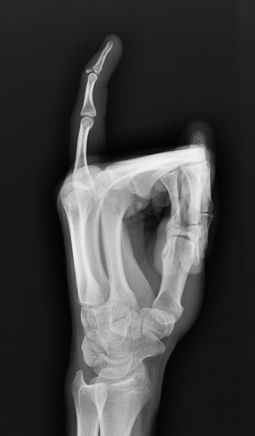
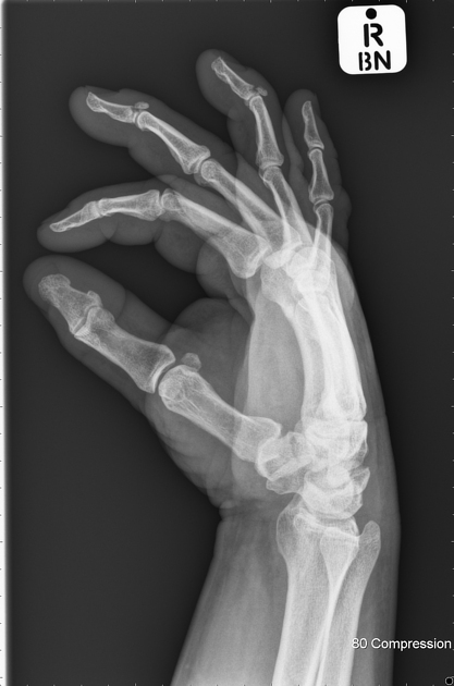
Plain radiograph
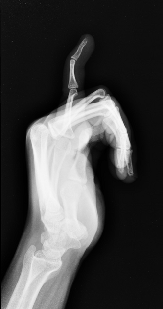
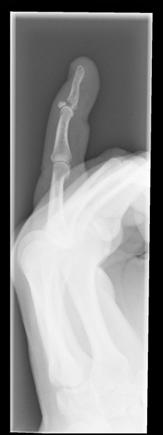
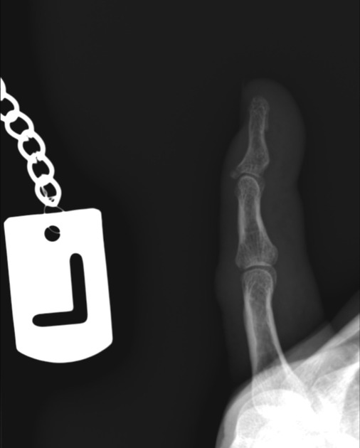
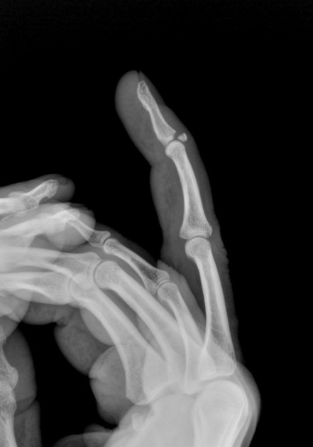
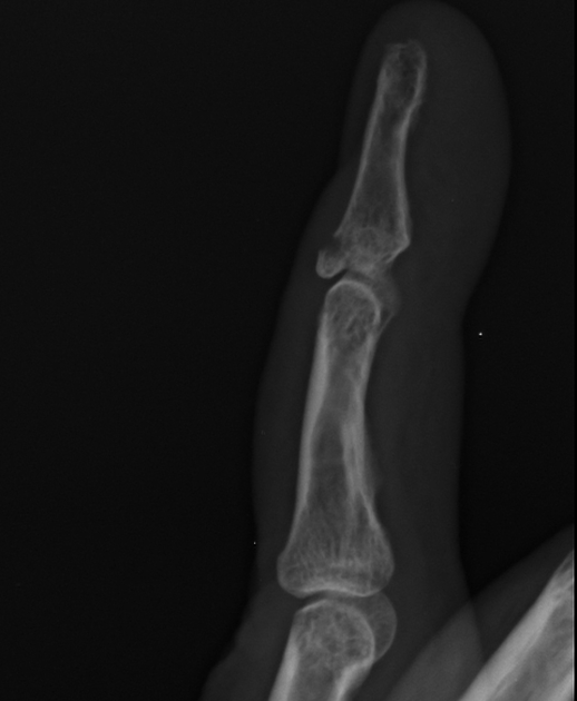
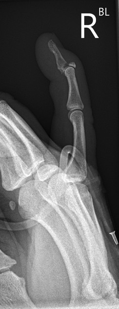
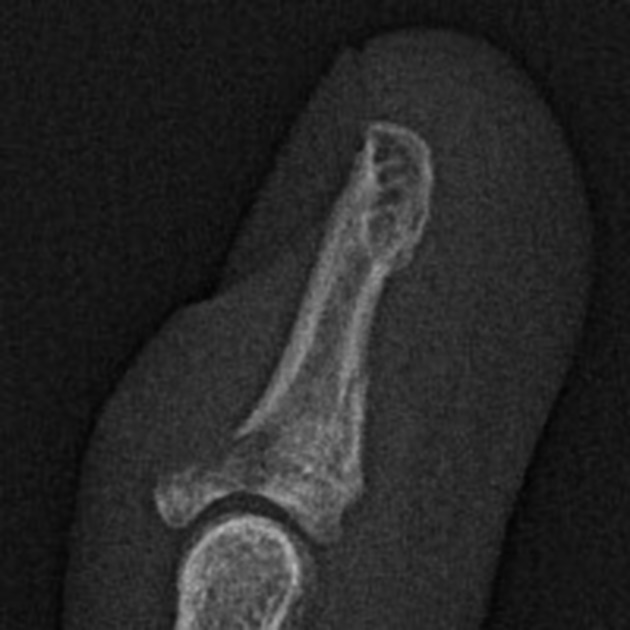
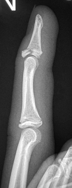
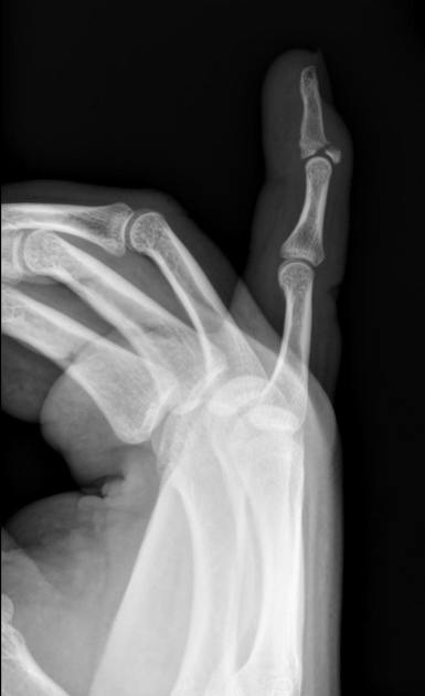
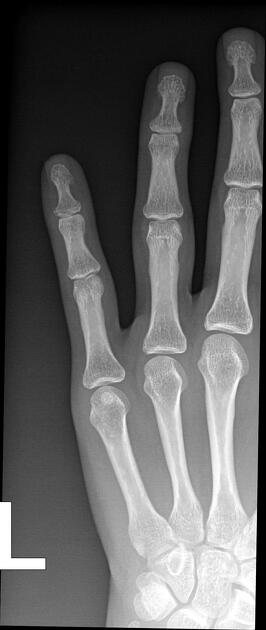
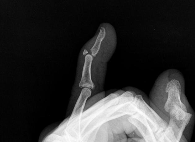
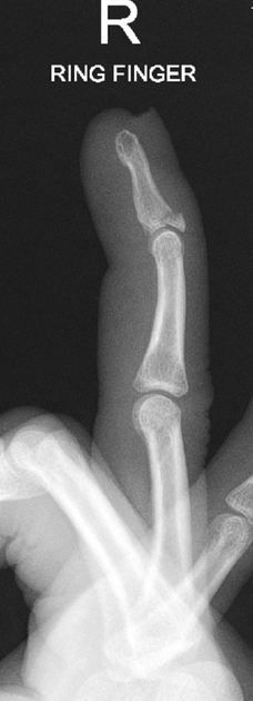
If there is a bony avulsion, a plain film will classically show a triangular avulsion fragment at the insertion of the common extensor tendon on the dorsal aspect of the distal phalanx at the DIP joint.
A high proportion of mallet finger injuries will present as isolated tendon injuries without any associated avulsions fractures known as a "mallet fracture" 5.
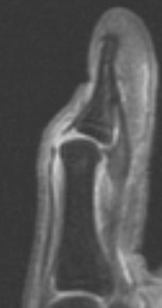
Ultrasound
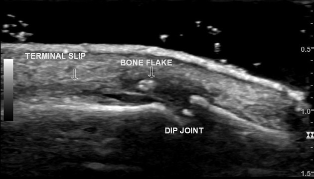
The findings on ultrasound include 6:
- loss of real-time movement of the tendon
- complete or partial extensor tendon tears
- fluid in the region of the extensor tendon insertion
- avulsion fracture
Treatment and prognosis
The preferred treatment for closed mallet injuries is non operative treatment, using a splint to maintain the DIP joint in extension or slight hyperextension - the proximal interphalangeal joint (PIP) is kept mobile. This positioning causes approximation of the injured tendon ends, which usually heals by scarring over time and restores extension 7. Non operative treatment would usually involve 6 weeks of full time splinting followed by 6 weeks of night splinting 8. It is also important to note that the DIP joint should be kept in full extension for the entire first 6 week period including times of hygiene.
It is uncommon for closed mallet finger injuries to require surgical intervention 5. Surgery is considered for avulsion fracture where the fragment is larger than 1/3 of the joint surface and there is more than 2 mm of displacement or there is volar subluxation of the distal phalanx which is not reducible in a splint.
The fragment may be pinned with Kirschner wire (either percutaneously or following open reduction) or indirectly reduced by "door stop" technique with the DIP flexed and stabilizing Kirschner wire placed through the middle phalanx 7. Post-operative complications, e.g. infection or need for further surgery, are common. The alternative is for fixation with a screw or hook plate if the fragments size will accommodate
Open injuries are generally surgically explored to evaluate for additional tendinous injury.
Complications
- the most common complication in mallet finger injuries are dorsal skin complications. This can include dorsal ulceration, nail deformities and maceration of the skin. These complications are prevalent in both operative and non operatively managed cases 8
- extensor lag
- untreated mallet finger or incomplete healing may progress to a swan neck deformity that will require surgical intervention 3,4,7
- development of secondary osteoarthritic changes 4
Related Radiopaedia articles
Fractures
-
fracture
- terminology
- fracture location
- diaphyseal fracture
- metaphyseal fracture
- physeal fracture
- epiphyseal fracture
- fracture types
- avulsion fracture
- articular surface injuries
- complete fracture
- incomplete fracture
- infraction
- compound fracture
- pathological fracture
- stress fracture
- fracture displacement
- fracture location
- fracture healing
- skull fractures
-
facial fractures
- fractures involving a single facial buttress
- alveolar process fractures
- frontal sinus fracture
- isolated zygomatic arch fractures
- mandibular fracture
- nasal bone fracture
- orbital blow-out fracture
- paranasal sinus fractures
- complex fractures
- dental fractures
- fractures involving a single facial buttress
-
spinal fractures
- classification (AO Spine classification systems)
-
cervical spine fracture classification systems
- AO classification of upper cervical injuries
- AO classification of subaxial injuries
- Anderson and D'Alonzo classification (odontoid fracture)
- Levine and Edwards classification (hangman fracture)
- Roy-Camille classification (odontoid process fracture )
- Allen and Ferguson classification (subaxial spine injuries)
- subaxial cervical spine injury classification (SLIC)
- thoracolumbar spinal fracture classification systems
- three column concept of spinal fractures (Denis classification)
- classification of sacral fractures
-
cervical spine fracture classification systems
- spinal fractures by region
- spinal fracture types
- classification (AO Spine classification systems)
- rib fractures
- sternal fractures
-
upper limb fractures
- classification
- Rockwood classification (acromioclavicular joint injury)
- AO classification (clavicle fracture)
- Neer classification (clavicle fracture)
- Neer classification (proximal humeral fracture)
- AO classification (proximal humeral fracture)
- AO/OTA classification of distal humeral fractures
- Milch classification (lateral humeral condyle fracture)
- Weiss classification (lateral humeral condyle fracture)
- Bado classification of Monteggia fracture-dislocations (radius-ulna)
- Mason classification (radial head fracture)
- Frykman classification (distal radial fracture)
- Mayo classification (scaphoid fracture)
- Hintermann classification (gamekeeper's thumb)
- Eaton classification (volar plate avulsion injury)
- Keifhaber-Stern classification (volar plate avulsion injury)
- upper limb fractures by region
- shoulder
- clavicular fracture
-
scapular fracture
- acromion fracture
- coracoid process fracture
- glenoid fracture
- humeral head fracture
- proximal humeral fracture
- humeral neck fracture
- arm
- elbow
- forearm
- wrist
-
carpal bones
- scaphoid fracture
- lunate fracture
- capitate fracture
- triquetral fracture
- pisiform fracture
- hamate fracture
- trapezoid fracture
- trapezium fracture
- hand
- shoulder
- classification
- lower limb fractures
- classification by region
- pelvis
- hip
- Pipkin classification (femoral head fracture)
- Garden classification (hip fracture)
- American Academy of Orthopedic Surgeons classification (periprosthetic hip fracture)
- Cooke and Newman classification (periprosthetic hip fracture)
- Johansson classification (periprosthetic hip fracture)
- Vancouver classification (periprosthetic hip fracture)
- femoral
- knee
- Schatzker classification (tibial plateau fracture)
- Meyers and McKeevers classification (anterior cruciate ligament avulsion fracture)
- tibia/fibula
- Watson-Jones classification (tibial tuberosity avulsion fracture)
- ankle
- foot
- Berndt and Harty classification (osteochondral lesions of the talus)
- Sanders CT classification (calcaneal fracture)
- Hawkins classification (talar neck fracture)
- Myerson classification (Lisfranc injury)
- Nunley-Vertullo classification (Lisfranc injury)
- pelvis and lower limb fractures by region
- pelvic fracture
- sacral fracture
- coccygeal fracture
-
hip
- acetabular fracture
- femoral head fracture
-
femoral neck fracture
- subcapital fracture
- transcervical fracture
- basicervical fracture
-
trochanteric fracture
- pertrochanteric fracture
- intertrochanteric fracture
- subtrochanteric fracture
- thigh
- mid-shaft fracture
- bisphosphonate-related fracture
- knee
- avulsion fractures
- Segond fracture
- reverse Segond fracture
- anterior cruciate ligament avulsion fracture
- posterior cruciate ligament avulsion fracture
- arcuate complex avulsion fracture (arcuate sign)
- biceps femoris avulsion fracture
- iliotibial band avulsion fracture
- semimembranosus tendon avulsion fracture
- Stieda fracture (MCL avulsion fracture)
- patellar fracture
- tibial plateau fracture
- avulsion fractures
- leg
- tibial tuberosity avulsion fracture
- tibial shaft fracture
- fibular shaft fracture
- Maisonneuve fracture
- ankle
- foot
- tarsal bones
- metatarsal bones
- phalanges
- classification by region
- terminology