
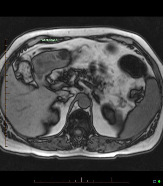
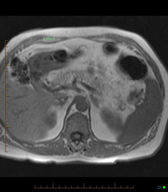
Pancreatic lipomatosis refers to fat accumulation in the pancreatic parenchyma. This finding is most often associated with obesity and aging and is the most common pathological condition involving the pancreas. Uneven fat deposition can simulate a pancreatic mass 1.
On this page:
Terminology
Synonyms include pancreatic lipomatosis, fatty pancreas and pancreatic steatosis 8,9. Non-alcoholic fatty pancreas (NAFP) excludes alcohol as a cause. Lipomatous pseudohypertrophy is characterized by pancreatic enlargement and may be a distinct entity 8.
The entity can be divided by etiology 8:
fatty replacement: death of acinar cells and subsequent replacement with adipocytes
fatty infiltration or nonalcoholic fatty pancreas disease: pancreatic accumulation of adipocytes in association with obesity and/or metabolic syndrome
Clinical presentation
The majority of pancreatic fatty deposits have no clinical significance. However, excessive fatty deposits may cause loss of pancreatic function, causing maldigestion of nutrients. This may lead to symptoms of chronic diarrhea, steatorrhea and weight loss, without any abdominal pain or signs of diabetes mellitus due to low insulin production 20. Symptomatic fatty pancreas is identified in several case reports 20.
Pathology
Subtypes
even pancreatic lipomatosis
-
uneven pancreatic lipomatosis 3,9
type 1a: preferential fatty replacement of the head, sparing the uncinate process and peribiliary region
type 1b: preferential fatty replacement of head, neck, and body, sparing the uncinate process and peribiliary region
type 2a: preferential fatty replacement of head, including uncinate process, and sparing the peribiliary region
type 2b: total fatty replacement of the pancreas except for the peribiliary region
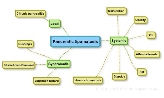
Etiology
Several etiologies are well established 8:
aging (most common degenerative lesion of the pancreas) 19
metabolic syndrome: obesity, dyslipidemia, diabetes mellitus
-
congenital syndromes
cystic fibrosis (most common cause in childhood)
carboxyl ester lipase mutation (maturity-onset diabetes of the young type 8) 10
Less established etiologies include the following 8:
-
chronic pancreatitis and/or pancreatic acinar atrophy due to a wide variety of insults including 19
viral infection
duct obstruction due to ligation, congenital stenosis, stones, or malignancy
excessive alcohol use 14
hemosiderosis / secondary hemochromatosis 16-18
-
drugs
corticosteroids 11
gemcitabine 12
cirrhosis related to chronic hepatitis B 13
-
malnutrition
Radiographic features
Ultrasound
On ultrasound, fatty pancreas has hyperechoic parenchyma 20.
CT
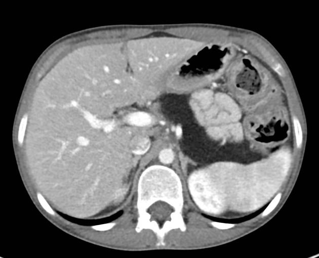
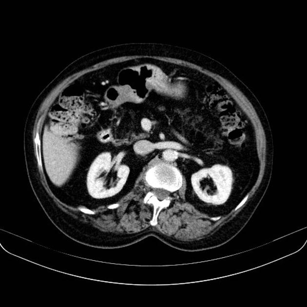
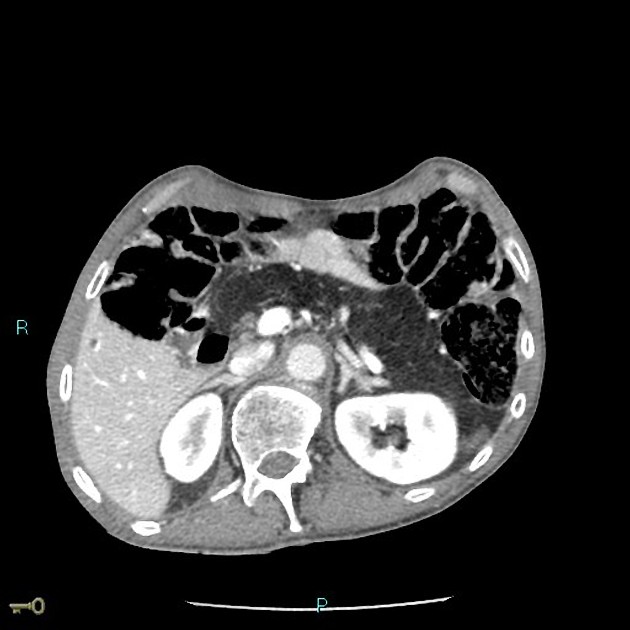
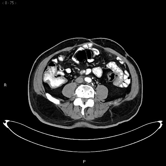
On CT scan, fatty pancreas has hypodense parenchyma 20. On unenhanced CT, the pancreas would show similar density to surrounding retroperitoneal fat (negative attenuation values). On contrast-enhanced CT, pancreatic parenchyma would show higher density (positive attenuation values) due to enhancement of pancreatic parenchyma between fatty infiltration areas 1.
Differential diagnosis
Imaging differential considerations include:
pancreatic agenesis: in pancreatic lipomatosis, the ducts remain preserved 3


 Unable to process the form. Check for errors and try again.