
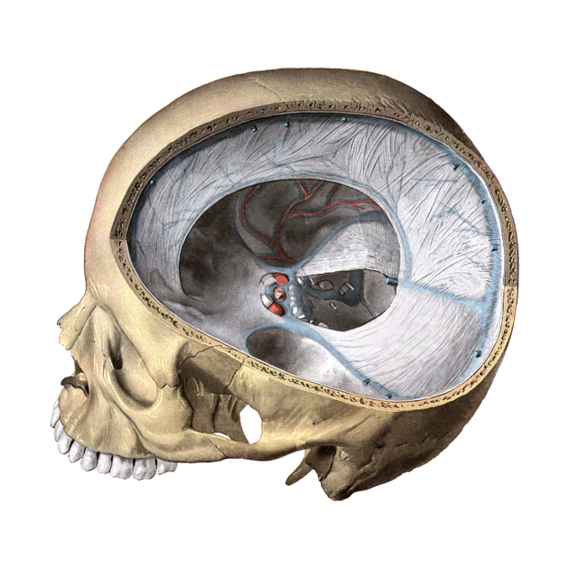
The tentorium cerebelli (plural: tentoria cerebellorum) is the second largest dural fold after the falx cerebri. It lies in the axial plane attached perpendicularly to the falx cerebri and divides the cranial cavity into supratentorial and infratentorial compartments 1. It has free and attached margins 2.
On this page:
Gross anatomy
The tentorium cerebelli is attached to the falx cerebri at its midline, and this attachment contains the straight sinus. This attachment is more superior than its anterolateral and posterolateral attachments giving it a "tented" appearance 1.
The anterior portion contains the tentorial notch (or tentorial incisure/incisura) which is a U-shaped (concave) free edge and extends anteriorly to attach to the anterior clinoid processes.1 The free edge forms a ridge of dura mater overlying the roof of the cavernous sinus from the superior petrosal sinus to its attachment on the anterior clinoid process.3 The deep tentorial notch surrounds the tentorial hiatus, which allows communication between the supratentorial and infratentorial compartments.
The anterolateral margin attaches to the superior border of the petrous part of the temporal bone. This margin lies inferior to the tentorial notch and extends anteriorly to attach to the posterior clinoid process and contains the superior petrosal sinus 1.
The posterolateral margin separates into the two layers of the dura and attaches to the edges of the transverse sulci of the occipital bone. As it extends anteriorly it attaches to the posteroinferior angles of the parietal bone 4. At its most posterior aspect, it is attached to the internal occipital protuberance 5 and posterolaterally contains the transverse sinus 4.
For blood supply and innervation, see dura.
Relations
superiorly: occipital lobe
inferiorly: cerebellum
anteriorly: midbrain, cerebral aqueduct
medially: extends over the anterior surface of the petrous part of the temporal bone forming Meckel cave
Radiographic features
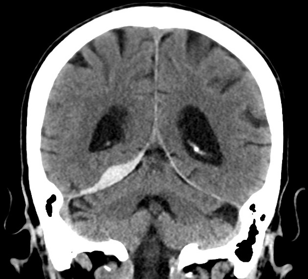
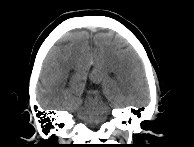
CT
-
non-contrast
appreciated best on coronal and sagittal views as a linear structure separating the occipital lobe from the cerebellum (or supratentorial compartment from the infratentorial compartment) 5
on coronal views is visible as being continuous with the falx cerebri
-
contrast-enhanced
on axial views tentorial margins best appreciated with IV contrast as uniform linear densities 5
Development
The tentorium cerebelli form from membranes derived from the cellular sheath of the notocord 6,7. These are divided into caudolateral and rostrolateral parts, that fuse in the midline posteriorly. Failure of normal fusion can lead to tentorial hypoplasia 7.


 Unable to process the form. Check for errors and try again.