
The shoulder, or shoulder joint, is the connection between the upper arm and the thorax. Comprising numerous ligamentous and muscular structures, the only actual bony articulations are the glenohumeral joint and the acromioclavicular joint (ACJ). The shoulder allows for an extensive range of motion due to the spheroid shape of the glenohumeral joint, but this (i.e. a large ball in a small socket) renders it prone to dislocation and other injuries.
On this page:
Gross anatomy
Location
The glenoid fossa of the scapula articulates with the anatomical head of the humerus as a synovial ball and socket joint. The glenoid fossa is deepened by the glenoid labrum.
The joint capsule attaches proximal to the glenoid fossa and further distally to the anatomical neck of the humerus. The capsule is looser inferiorly to allow for tightening during the abduction.
Movements
flexion: anterior fibers of deltoid, coracobrachialis, biceps brachii
extension: posterior fibers of deltoid, teres minor, teres major
abduction: supraspinatus, deltoid, trapezius
adduction: pectoralis major, subscapularis, teres major, latissimus dorsi
internal rotation: teres major, latissimus dorsi, pectoralis major
external rotation: infraspinatus, teres minor
Bursae
Numerous bursae are associated with the shoulder joint, which include the subacromial-subdeltoid and subcoracoid bursae.
Ligaments
superior, middle and inferior glenohumeral ligaments, from their attachments along the anterior surface of the glenoid fossa extending further inferiorly
transverse humeral ligament (holds long head of biceps down as it runs in the intertubercular groove)
long head of biceps (from supraglenoid tubercle) is intracapsular, running in the superior portion of the joint
long head of triceps at its origin (from infraglenoid tubercle) also supports the joint inferiorly
also reinforced by important extrinsic ligaments including the acromioclavicular, coracoacromial and coracoclavicular ligaments
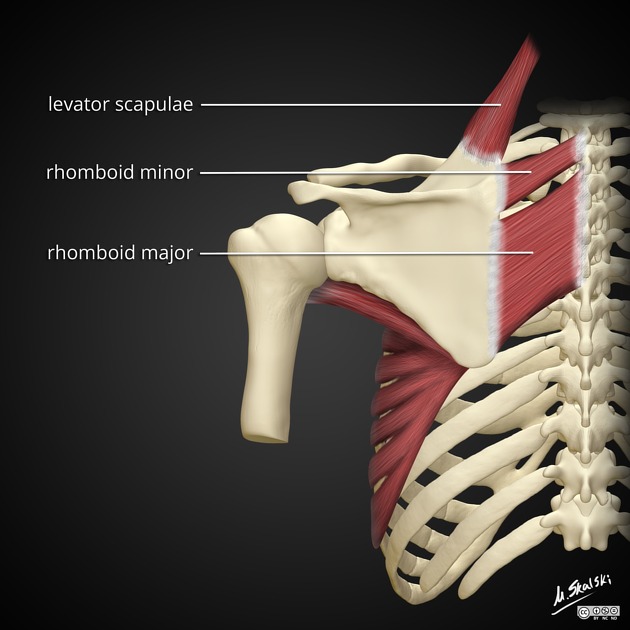
Tendons
The shoulder joint is reinforced by the rotator cuff muscles/tendons:
supraspinatus: from supraspinous fossa to superior facet on greater tubercle
infraspinatus: from infraspinous fossa to middle facet on greater tubercle
teres minor: from inferolateral border of the scapula to the inferior facet on the greater tubercle
subscapularis: from the subscapular fossa to the lesser tubercle
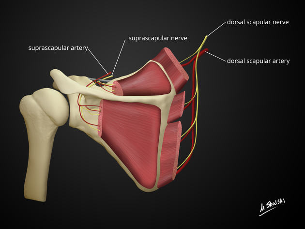
Arterial supply
Arterial supply is from the branches of the scapular anastomosis but primarily from the axillary artery:
Venous drainage
Veins with corresponding names, accompany the arteries, and drain the shoulder via a periscapular venous plexus.
Innervation
Multiple articular branches are derived from several nerves (Hilton's law):
Related pathology
-
signs
-
traumatic
-
humeral head injuries
Reverse Hill-Sachs defect (McLaughlin lesion)
-
metabolic
-
degenerative
-
others
adhesive capsulitis of the shoulder (frozen shoulder)
anterosuperior inner impingement


 Unable to process the form. Check for errors and try again.