
Normal contours of the cardiomediastinum on chest radiography
Last revised by Bálint Botz
on 28 Jun 2021
Citation, DOI, disclosures and article data
Citation:
Gaillard F, Botz B, Dixon A, et al. Normal contours of the cardiomediastinum on chest radiography. Reference article, Radiopaedia.org (Accessed on 16 Feb 2025) https://doi.org/10.53347/rID-8469
Permalink:
rID:
8469
Article created:
Disclosures:
At the time the article was created Frank Gaillard had no recorded disclosures.
View Frank Gaillard's current disclosures
Last revised:
28 Jun 2021,
Bálint Botz
Disclosures:
At the time the article was last revised Bálint Botz had no recorded disclosures.
View Bálint Botz's current disclosures
Revisions:
11 times, by
9 contributors -
see full revision history and disclosures
Sections:
Tags:
Synonyms:
- Normal cardiomediastinal outline
A detailed understanding of the structures that make up the normal contours of the heart and mediastinum (cardiomediastinal contour) on chest radiography is essential if abnormalities are to be detected.
Frontal view (PA/AP)
Right cardiomediastinal contour
From superior to inferior:
-
right paratracheal stripe
- seen in two thirds of normal films 1
- made up of right brachiocephalic vein and SVC
- arch of the azygos vein
- ascending aorta in older individuals often projects to the right of the SVC
- superior vena cava (SVC)
- right atrium
- inferior vena cava (IVC)
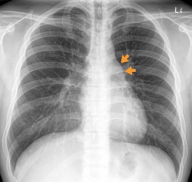
Left cardiomediastinal contour
From superior to inferior:
-
left paratracheal stripe
- made up of left common carotid artery, left subclavian artery and the left jugular vein
- aortic arch +/- aortic nipple (left superior intercostal vein)
- pulmonary trunk
- auricle of left atrium
- left ventricle
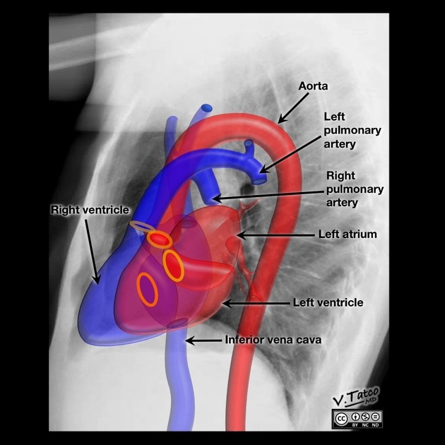
Lateral view
Anterior cardiomediastinal contour
From superior to inferior:
-
superior mediastinum
- great vessels
- thymus
- ascending aorta
- right ventricular outflow tract
- right ventricle
Posterior cardiomediastinal contour
From superior to inferior:
- left atrium and pulmonary veins
- left ventricle
- inferior vena cava
Quiz questions
References
- 1. Collins J, Stern EJ. Chest radiology, the essentials. Lippincott Williams & Wilkins. (2007) ISBN:0781763142. Read it at Google Books - Find it at Amazon
- 2. Haller JO, Slovis TL, Joshi A. Pediatric radiology. Springer Verlag. (2005) ISBN:3540213546. Read it at Google Books - Find it at Amazon
Incoming Links
Articles:
- Right upper lobe collapse in the exam
- Cardiac chamber enlargement
- Chest radiograph
- Moguls of the heart
- Chest (supine view)
- Ascending aorta
- Chest curriculum
- Enlargement of the cardiac silhouette
- Right middle lobe collapse
- Right upper lobe collapse
- Left lower lobe collapse
- Left upper lobe collapse
- Assessment of cardiomediastinal contours on chest x-ray (approach)
- Chest (lateral view)
- Posterior mediastinal mass in the exam
- Azygos vein
- Traumatic aortic injury in the exam
- Left atrial enlargement
- Chest (AP erect view)
- Atrial escape
Cases:
- Pulmonary agenesis and dextrocardia
- Normal chest radiograph - pediatric
- Left ventricular enlargement on chest radiography (illustration)
- Left atrial enlargement on chest radiography (illustration)
- Right ventricular enlargement on chest radiography (illustration)
- Right atrial enlargement on chest radiography (illustration)
- Moguls of the heart (illustration)
- Left lower lobe collapse
- Retrosternal goitre
- Right middle lobe collapse
- Right lower lobe collapse
- Left upper lobe collapse
- Cardiomegaly
- Left upper lobe collapse
- Right middle lobe consolidation - patchy
- Right middle lobe consolidation - patchy
- Cardiomediastinal outlines on chest x-ray
- Pericardial effusion - water bottle sign
Multiple choice questions:
Related articles: Chest
- imaging techniques
-
chest radiograph
- radiography
-
approach
- ABCDE
- ABCDEFGHI
- congenital heart disease
- medical devices in the thorax
- common lines and tubes
- nasogastric tubes
- endotracheal tubes
- central venous catheters
- esophageal temperature probe
- tracheostomy tube
- pleural catheters
- cardiac conduction devices
- prosthetic heart valve
- review areas
-
airspace opacification
- differential diagnoses of airspace opacification
- lobar consolidation
-
atelectasis
- mechanism-based
- morphology-based
- lobar lung collapse
- chest x-ray in the exam setting
- cardiomediastinal contour
- chest radiograph zones
- tracheal air column
- fissures
- normal chest x-ray appearance of the diaphragm
- nipple shadow
-
lines and stripes
- anterior junction line
- posterior junction line
- right paratracheal stripe
- left paratracheal stripe
- posterior tracheal stripe/tracheo-esophageal stripe
- posterior wall of bronchus intermedius
- right paraspinal line
- left paraspinal line
- aortic-pulmonary stripe
- aortopulmonary window
- azygo-esophageal recess
- spaces
- signs
- air bronchogram
- big rib sign
- Chang sign
- Chen sign
- coin lesion
- continuous diaphragm sign
- dense hilum sign
- double contour sign
- egg-on-a-string sign
- extrapleural sign
- finger in glove sign
- flat waist sign
- Fleischner sign
- ginkgo leaf sign
- Golden S sign
- Hampton hump
- haystack sign
- hilum convergence sign
- hilum overlay sign
- Hoffman-Rigler sign
- holly leaf sign
- incomplete border sign
- juxtaphrenic peak sign
- Kirklin sign
- medial stripe sign
- melting ice cube sign
- more black sign
- Naclerio V sign
- Palla sign
- pericardial fat tag sign
- Shmoo sign
- silhouette sign
- snowman sign
- spinnaker sign
- steeple sign
- straight left heart border sign
- third mogul sign
- tram-track sign
- walking man sign
- water bottle sign
- wave sign
- Westermark sign
- HRCT
-
chest radiograph
- airways
- bronchitis
- small airways disease
-
bronchiectasis
- broncho-arterial ratio
- related conditions
- differentials by distribution
- narrowing
-
tracheal stenosis
- diffuse tracheal narrowing (differential)
-
bronchial stenosis
- diffuse airway narrowing (differential)
-
tracheal stenosis
- diverticula
- pulmonary edema
-
interstitial lung disease (ILD)
- Anti-Jo-1 antibody-positive interstitial lung disease
- drug-induced interstitial lung disease
-
hypersensitivity pneumonitis
- acute hypersensitivity pneumonitis
- subacute hypersensitivity pneumonitis
- chronic hypersensitivity pneumonitis
- etiology
- bird fancier's lung: pigeon fancier's lung
- farmer's lung
- cheese workers' lung
- bagassosis
- mushroom worker’s lung
- malt worker’s lung
- maple bark disease
- hot tub lung
- wine maker’s lung
- woodsman’s disease
- thatched roof lung
- tobacco grower’s lung
- potato riddler’s lung
- summer-type pneumonitis
- dry rot lung
- machine operator’s lung
- humidifier lung
- shower curtain disease
- furrier’s lung
- miller’s lung
- lycoperdonosis
- saxophone lung
-
idiopathic interstitial pneumonia (mnemonic)
- acute interstitial pneumonia (AIP)
- cryptogenic organizing pneumonia (COP)
- desquamative interstitial pneumonia (DIP)
- non-specific interstitial pneumonia (NSIP)
- idiopathic pleuroparenchymal fibroelastosis
- lymphoid interstitial pneumonia (LIP)
- respiratory bronchiolitis–associated interstitial lung disease (RB-ILD)
- usual interstitial pneumonia / idiopathic pulmonary fibrosis (UIP/IPF)
-
pneumoconioses
- fibrotic
- non-fibrotic
-
lung cancer
-
non-small-cell lung cancer
-
adenocarcinoma
- pre-invasive tumors
- minimally invasive tumors
- invasive tumors
- variants of invasive carcinoma
- described imaging features
- adenosquamous carcinoma
- large cell carcinoma
- primary sarcomatoid carcinoma of the lung
- squamous cell carcinoma
- salivary gland-type tumors
-
adenocarcinoma
- pulmonary neuroendocrine tumors
- preinvasive lesions
-
lung cancer invasion patterns
- tumor spread through air spaces (STAS)
- presence of non-lepidic patterns such as acinar, papillary, solid, or micropapillary
- myofibroblastic stroma associated with invasive tumor cells
- pleural invasion
- vascular invasion
- tumors by location
- benign neoplasms
- pulmonary metastases
- lung cancer screening
- lung cancer staging
-
non-small-cell lung cancer


 Unable to process the form. Check for errors and try again.