
The ophthalmic artery is a branch of the supraclinoid (C6) segment of the internal carotid artery.
Gross anatomy
Origin
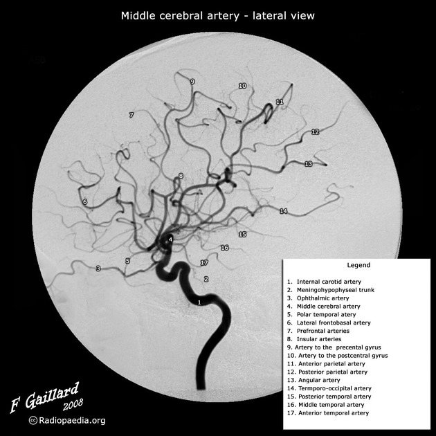
The ophthalmic artery arises medial to the anterior clinoid process as the internal carotid artery exits the cavernous sinus. It originates from the antero- or supero-medial surface of the internal carotid artery.
Course
The ophthalmic artery passes into the orbit via the optic canal. It has numerous branches which are often grouped into those that supply the orbital content and those that supply the globe and related structures.
Termination
The two terminal branches of the ophthalmic artery are the supratrochlear artery and the dorsal nasal artery.
Branches
Orbital group
supratrochlear artery (frontal artery)
Ocular group
central retinal artery: although rarely seen, the choroidal blush should be seen on all angiograms
-
long posterior ciliary arteries (supply the anterior part of the choroid and ciliary body and iris) 4
short posterior ciliary arteries (supply the posterior part of the choroid) 4
muscular artery
A useful mnemonic to remember the branches of the ophthalmic artery is:
Variant anatomy
Embryologically, the orbit has dual supply: from the supraorbital branch (which later becomes the middle meningeal artery) and from the ophthalmic artery. As a result there can be substantial variation 1,2:
-
single communicating branch between the ophthalmic and middle meningeal artery
present in up to 50% of individuals
most frequently passes through the superior orbital fissure
known as sphenoidal artery or recurrent meningeal artery or orbital branch of the middle meningeal branch
-
multiple communicating branches
present in up to 15% of individuals
pass through additional small foramen (or multiple foramina) lateral to the superior orbital fissure are present (foramen of Hyrtl or the meningo-orbital foramen)
-
meningo-ophthalmic artery: regression of proximal ophthalmic artery and entire orbit is supplied by the middle meningeal artery (4%) 4
enters the orbit via the superior orbital fissure or via its own canal.
middle meningeal artery can arise from the ophthalmic artery
ophthalmic artery origin from the middle cerebral artery or posterior communicating artery
-
persistent dorsal ophthalmic artery (cavernous segment origin)
present in 10% of individuals 4
These variants are important to recognise during endovascular thereapy. For example, embolisation of a tumour supplied by the external carotid artery (e.g. meningioma or haemangiopericytoma) can result in blindness.


 Unable to process the form. Check for errors and try again.