
Cavernous sinus
Citation, DOI, disclosures and article data
At the time the article was created Frank Gaillard had no recorded disclosures.
View Frank Gaillard's current disclosuresAt the time the article was last revised Craig Hacking had the following disclosures:
- Philips Australia, Paid speaker at Philips Spectral CT events (ongoing)
These were assessed during peer review and were determined to not be relevant to the changes that were made.
View Craig Hacking's current disclosures- Cavernous sinuses
The cavernous sinuses are paired dural venous sinuses.
Gross anatomy
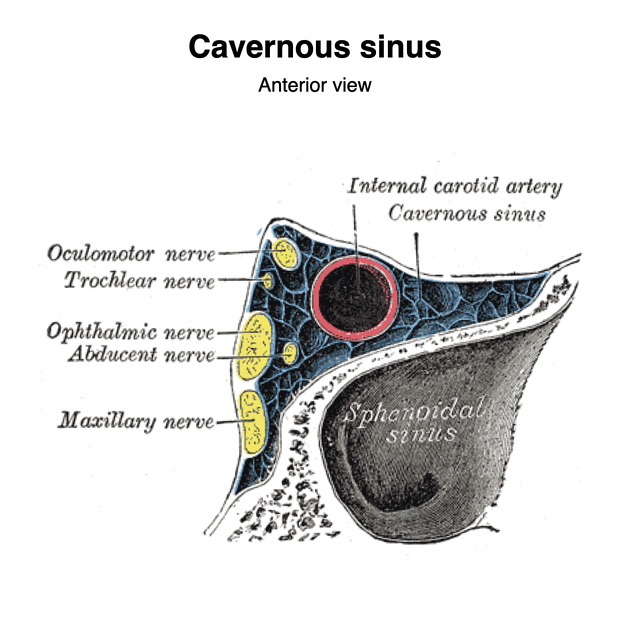
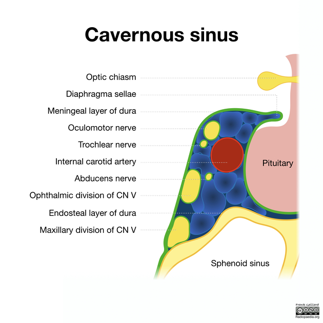
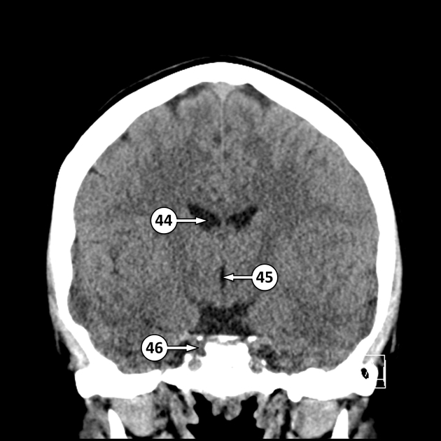
The cavernous sinus is located on either side of the pituitary fossa and body of the sphenoid bone. It is most easily thought of as existing between the endosteal and meningeal layers of the dura although some additional complexity is present 5. It spans from the apex of the orbit to the apex of the petrous temporal bone. Unlike other dural venous sinuses, it is divided by numerous fibrous septa into a series of small caves, which is where its name is derived from. The normal lateral wall should be either straight or concave.
Boundaries
- roof: fold of dura mater attached to the anterior and middle clinoid processes
- anterior wall: medial end of the superior orbital fissure
- posterior wall: petrous apex
- medial wall: endosteum overlying the body of the sphenoid bone
- lateral wall: dura mater from the ridge of the roof to the floor of the middle cranial fossa
- floor: endosteum overlying the base of the greater wing of the sphenoid bone
Relations
- superiorly: middle cerebral artery, optic chiasm
- anteriorly: apex of the orbit
- posteriorly: cerebral peduncle
- medially: pituitary fossa, pituitary gland
- laterally: temporal lobe (medial surface), Meckel's cave (posteroinferiorly)
- inferiorly: sphenoid sinus
Dural anatomy
The lateral wall of the cavernous sinus is primarily formed by the continuation of the meningeal layer of the dura, flowing medially up from the floor of the middle cranial fossa, over the cavernous sinus, to the clinoid processes before forming the diaphragma sella. From here, the meningeal layer passes downwards to surround the pituitary gland, thus forming the medial wall of the cavernous sinus. In contrast, the floor of the cavernous sinus is formed by the endosteal layer of the meninges (actually just periosteum) that covers the sphenoid bone and passes medially across the midline below the pituitary gland, separated from the aforementioned meningeal layer by the intercavernous sinus 5.
The nerves in the lateral wall of the cavernous sinus are surrounded by thin connective tissue and thus, the lateral wall of the cavernous sinus has two layers; a thick meningeal layer and a much thinner translucent layer surrounding the cranial nerves 5,6. This thin layer is variably described as being meningeal in origin 6, or continuous with the endosteal layer 5.
Vascular connections
It receives venous blood from:
- inferior and superior ophthalmic veins
- intercavernous sinus
- sphenoparietal sinus
- superficial middle cerebral vein
- occasionally
- central retinal vein
- a frontal tributary of the middle meningeal vein
Drainage of the cavernous sinus is via:
- superior petrosal sinus to the transverse sinus
- inferior petrosal sinus directly to the jugular bulb
- venous plexus on the internal carotid artery (ICA) to the clival (basilar) venous plexuses
- emissary veins passing through the
- foramen Vesalii
- foramen ovale: communicates between the cavernous sinus and pterygoid venous plexus
- foramen lacerum
Depending on relative pressures the superior ophthalmic veins either drain to or from the cavernous sinus.
Additionally, the cavernous sinuses connect to each other via the intercavernous sinuses.
Contents
These can be remembered with the mnemonic O TOM CAT.
Nerves
The cavernous sinus transmits multiple cranial nerves to the superior orbital fissure and foramen rotundum. These are:
- in the lateral wall from superior to inferior
- oculomotor nerve (CN III)
- trochlear nerve (CN IV)
-
trigeminal nerve (CN V)
- ophthalmic division
- maxillary division: within the very inferolateral aspect of the cavernous sinus wall or even outside the sinus rather than truly within it 4
- traversing the sinus
- abducens nerve (CN VI): inferolateral to the internal carotid artery
Artery
The internal carotid artery enters the posterior inferior aspect of the sinus and bends upon itself as the carotid siphon (cavernous segment - C4). Two branches arise from this segment: meningohypophyseal trunk and inferolateral trunk.
The artery is surrounded by a plexus of sympathetic nerves from the superior cervical ganglion.
Fat
Fatty deposits may be present within the cavernous sinus, especially in obese patients or in those who are taking corticosteroids 3.
ADVERTISEMENT: Supporters see fewer/no ads
Related pathology
Quiz questions
Question 2398
An MRI of a patient with left facial numbness and ophthalmoplegia shows a T2 intermediate signal intensity mass in the left cavernous sinus which exhibits homogeneous enhancement and extends into Meckel’s cave and the orbital apex. The flow void of the left internal carotid artery is mildly narrowed. CT shows mild thickening and sclerosis of the lateral wall of the left sphenoid sinus.
What is the most likely diagnosis?
References
- 1. Standring S, Gray H. Gray's anatomy, the anatomical basis of clinical practice. Churchill Livingstone. (2008) ISBN:0443066841. Read it at Google Books - Find it at Amazon
- 2. Safdieh JE. Netter's Concise Neuroanatomy. Saunders. (2007) ISBN:1933247223. Read it at Google Books - Find it at Amazon
- 3. Razek AA, Castillo M. Imaging lesions of the cavernous sinus. AJNR Am J Neuroradiol. 2009;30 (3): 444-52. doi:10.3174/ajnr.A1398 - Pubmed citation
- 4. Tubbs RS, Hill M, May WR, Middlebrooks E, Kominek SZ, Marchase N, Shoja MM, Loukas M, Oakes WJ. Does the maxillary division of the trigeminal nerve traverse the cavernous sinus? An anatomical study and review of the literature. (2008) Surgical and radiologic anatomy : SRA. 30 (1): 37-40. doi:10.1007/s00276-007-0280-7 - Pubmed
- 5. Yasuda A, Campero A, Martins C, Rhoton A, de Oliveira E, Ribas G. Microsurgical Anatomy and Approaches to the Cavernous Sinus. Neurosurgery. 2008;62(6):SHC1240-63. doi:10.1227/01.neu.0000333790.90972.59 - Pubmed
- 6. Kawase T, van Loveren H, Keller J, Tew J. Meningeal Architecture of the Cavernous Sinus: Clinical and Surgical Implications. Neurosurgery. 1996;39(3):527-35. doi:10.1097/00006123-199609000-00019 - Pubmed
Incoming Links
- Dorello canal
- Inferior petrosal sinus
- Dura mater
- Pituitary fossa
- Internal carotid artery venous plexus of Rektorzik
- Zurich pituitary score
- Pterygopalatine fossa
- Suboccipital cavernous sinus
- Superior ophthalmic vein
- Foramen Vesalii
- Ophthalmic artery
- Abducens nerve
- Vein of Galen aneurysmal malformation
- Tuberculous pachymeningitis
- Superior petrosal sinus
- Superior orbital fissure
- Blunt cerebrovascular injury
- Dural arteriovenous fistula
- Cavernous sinus contents (mnemonic)
- Cavernous sinus syndrome
- Tolosa-Hunt syndrome
- Caroticocavernous fistula
- Fat in cavernous system
- Bilateral cavernous segments internal carotid artery aneurysms
- Pituitary macroadenoma with apoplexy
- Trigeminal amyloidoma
- Normal MRI orbits
- Idiopathic intracranial hypertension
- Bilateral indirect carotid cavernous fistula
- Fat in normal cavernous sinus
- Meningioma - cavernous sinus
- Cavernous sinus fat
- Cavernous sinus (Gray's illustration)
- Bilateral plexiform neurofibromas of the trigeminal and facial nerves - NF1
- Internal carotid artery aneurysm
- Idiopathic intracranial hypertension
- Cavernous sinus thrombosis
- Cavernous sinus arachnoid cyst
- En plaque meningioma with associated CPA / frontal convexity meningiomas
- Tolosa-Hunt syndrome
Related articles: Anatomy: Brain
-
brain
- grey matter
- white matter
-
cerebrum
-
cerebral hemisphere (telencephalon)
- cerebral lobes and gyri
- frontal lobe
- parietal lobe
-
occipital lobe
- occipital pole
- lingual gyrus
- fusiform gyrus (Brodmann area 37)
- calcarine (visual) cortex
- cuneus
- temporal lobe
- basal forebrain
- limbic system
- insula
-
cerebral sulci and fissures (A-Z)
- calcarine fissure
- callosal sulcus
- central (Rolandic) sulcus
- cingulate sulcus
- collateral sulcus
- inferior frontal sulcus
- inferior occipital sulcus
- inferior temporal sulcus
- interhemispheric fissure
- intraparietal sulcus
- lateral (Sylvian) sulcus
- lateral occipital sulcus
- marginal sulcus
- occipitotemporal sulcus
- olfactory sulcus
- paracentral sulcus
- paraolfactory sulcus
- parieto-occipital fissure
- posterior parolfactory sulcus
- precentral sulcus
- preoccipital notch
- postcentral sulcus
- rhinal sulcus
- rostral sulcus
- subparietal sulcus
- superior frontal sulcus
- superior occipital sulcus
- superior temporal sulcus
- cortical histology
- cerebral lobes and gyri
- white matter tracts
- deep grey matter
-
pituitary gland
- posterior pituitary and stalk (part of diencephalon)
- anterior pituitary
- inferior hypophyseal arterial circle
- diencephalon
-
cerebral hemisphere (telencephalon)
-
brainstem
- midbrain (mesencephalon)
- pons (part of metencephalon)
- medulla oblongata (myelencephalon)
- white matter
-
grey matter
- non-cranial nerve
-
cranial nerve nuclei
- oculomotor nucleus
- Edinger-Westphal nucleus
- trochlear nucleus
- motor nucleus of CN V
- mesencephalic nucleus of CN V
- main sensory nucleus of CN V
- spinal nucleus of CN V
- abducent nucleus
- facial nucleus
- superior salivatory nucleus
- cochlear nuclei
- vestibular nuclei
- inferior salivatory nucleus
- solitary tract nucleus
- ambiguus nucleus
- dorsal vagal motor nucleus
- hypoglossal nucleus
-
cerebellum (part of metencephalon)
- vermis
- cerebellar hemisphere
- cerebellar peduncles
- cranial meninges (meninx primitiva)
- CSF spaces
-
cranial nerves (mnemonic)
- olfactory nerve (CN I)
- optic nerve (CN II)
- oculomotor nerve (CN III)
- trochlear nerve (CN IV)
- trigeminal nerve (CN V) (mnemonic)
- abducens nerve (CN VI)
- facial nerve (CN VII) (segments mnemonic | branches mnemonic)
-
vestibulocochlear nerve (CN VIII)
- vestibular ganglion (Scarpa's ganglion)
- glossopharyngeal nerve (CN IX)
- vagus nerve (CN X)
- spinal accessory nerve (CN XI)
- hypoglossal nerve (CN XII)
- functional neuroanatomy
- CNS development
- cerebral vascular supply
- arteries
- vascular territories
-
circle of Willis
- internal carotid artery (ICA) (segments)
- vertebral artery
-
normal variants
- intracranial arterial fenestration
- internal carotid artery (ICA)
- anterior cerebral artery (ACA)
- middle cerebral artery (MCA)
- posterior cerebral artery (PCA)
- basilar artery
- persistent carotid-vertebrobasilar artery anastomoses (mnemonic)
- vertebral artery
- ophthalmic artery
-
cerebral venous system
-
dural venous sinuses
- basilar venous plexus
- cavernous sinus (mnemonic)
- clival diploic veins
- inferior petro-occipital vein
- inferior petrosal sinus
- inferior sagittal sinus
- intercavernous sinus
- internal carotid artery venous plexus of Rektorzik
- jugular bulb
- marginal sinus
- occipital sinus
- sigmoid sinus
- sphenoparietal sinus
- straight sinus
- superior petrosal sinus
- superior sagittal sinus
- torcula herophili
- transverse sinus
-
cerebral veins
-
superficial veins of the brain
- superior cerebral veins (superficial cerebral veins)
- inferior cerebral veins
- superficial middle cerebral vein
- superior anastomotic vein (of Trolard)
- inferior anastomotic vein (of Labbe)
-
superficial veins of the brain
-
deep veins of the brain
- great cerebral vein (of Galen)
- venous circle of Trolard
- normal variants
-
dural venous sinuses
- arteries
- glymphatic pathway
Related articles: Anatomy: Head and neck
- skeleton of the head and neck
-
cranial vault
- scalp (mnemonic)
- fontanelle
-
sutures
- calvarial
- facial
- frontozygomatic suture
- frontomaxillary suture
- frontolacrimal suture
- frontonasal suture
- temporozygomatic suture
- zygomaticomaxillary suture
- parietotemporal suture (parietomastoid suture)
- occipitotemporal suture (occipitomastoid suture)
- sphenofrontal suture
- sphenozygomatic suture
- spheno-occipital suture (not a true suture)
- lacrimomaxillary suture
- nasomaxillary suture
- internasal suture
- basal/internal
- skull landmarks
- frontal bone
- temporal bone
- parietal bone
- occipital bone
- skull base (foramina)
-
facial bones
- midline single bones
- paired bilateral bones
- cervical spine
- hyoid bone
- laryngeal cartilages
-
cranial vault
- muscles of the head and neck
- muscles of the tongue (mnemonic)
- muscles of mastication
-
facial muscles
- epicranius muscle
- circumorbital and palpebral muscles
- nasal muscles
-
buccolabial muscles
- elevators, retractors and evertors of the upper lip
- levator labii superioris alaeque nasalis muscle
- levator labii superioris muscle
- zygomaticus major muscle
- zygomaticus minor muscle
- levator anguli oris muscle
- malaris muscle
- risorius muscle
- depressors, retractors and evertors of the lower lip
- depressor labii inferioris muscle
- depressor anguli oris muscle
- mentalis muscle
- compound sphincter
-
orbicularis oris muscle
- incisivus labii superioris muscle
- incisivus labii inferioris muscle
-
orbicularis oris muscle
- muscle of mastication
- modiolus
- elevators, retractors and evertors of the upper lip
- muscles of the middle ear
- orbital muscles
- muscles of the soft palate
- pharyngeal muscles
- suprahyoid muscles
- infrahyoid muscles
- intrinsic muscles of the larynx
- muscles of the neck
- platysma muscle
- longus colli muscle
- longus capitis muscle
- scalenus anterior muscle
- scalenus medius muscle
- scalenus posterior muscle
- scalenus pleuralis muscle
- sternocleidomastoid muscle
-
suboccipital muscles
- rectus capitis posterior major muscle
- rectus capitis posterior minor muscle
- obliquus capitis superior muscle
- obliquus capitis inferior muscle
- accessory muscles of the neck
- deep cervical fascia
-
deep spaces of the neck
- anterior cervical space
- buccal space
- carotid space
- danger space
- deep cervical fascia
- infratemporal fossa
- masticator space
- parapharyngeal space
- stylomandibular tunnel
- parotid space
- pharyngeal (superficial) mucosal space
- perivertebral space
- posterior cervical space
- pterygopalatine fossa
- retropharyngeal space
- suprasternal space (of Burns)
- visceral space
- surgical triangles of the neck
- orbit
- ear
- paranasal sinuses
- upper respiratory tract
- viscera of the neck
- blood supply of the head and neck
-
arterial supply
-
common carotid artery
- carotid body
- carotid bifurcation
- subclavian artery
- variants
-
common carotid artery
- venous drainage
-
arterial supply
- innervation of the head and neck
-
cranial nerves
- olfactory nerve (CN I)
- optic nerve (CN II)
- oculomotor nerve (CN III)
- trochlear nerve (CN IV)
-
trigeminal nerve (CN V) (mnemonic)
- trigeminal ganglion
- ophthalmic division
- maxillary division
- mandibular division
- abducens nerve (CN VI)
- facial nerve (CN VII)
-
vestibulocochlear nerve (CN VIII)
- vestibular ganglion (Scarpa's ganglion)
- glossopharyngeal nerve (CN IX)
- vagus nerve (CN X)
- (spinal) accessory nerve (CN XI)
- hypoglossal nerve (CN XII)
- parasympathetic ganglia of the head and neck
- cervical sympathetic ganglia
- greater occipital nerve
- third occipital nerve
-
cervical plexus
- muscular branches
- longus capitis
- longus colli
- scalenes
- geniohyoid
- thyrohyoid
-
ansa cervicalis
- omohyoid (superior and inferior bellies separately)
- sternothyroid
- sternohyoid
- phrenic nerve
- contribution to the accessory nerve (CN XI)
- cutaneous branches
- muscular branches
- brachial plexus
- pharyngeal plexus
-
cranial nerves
- lymphatic drainage of the head and neck
- embryological development of the head and neck


 Unable to process the form. Check for errors and try again.