
Right atrial (RA) enlargement is less common, and harder to delineate on chest radiograph, than left atrial (LA) enlargement.
On this page:
Pathology
Etiology
Enlargement of the right atrium (RA) can result from a number of conditions, including:
-
raised right ventricular pressures
-
valvular disease
Clinical presentation
ECG
-
amplitude of P wave in lead II >2.5 mm
often demonstrate "peaked" morphology
a.k.a. P pulmonale 7
initial positive deflection of P wave in lead V1 >1.5 mm
Radiographic features
Plain radiograph
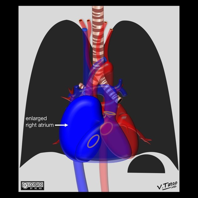
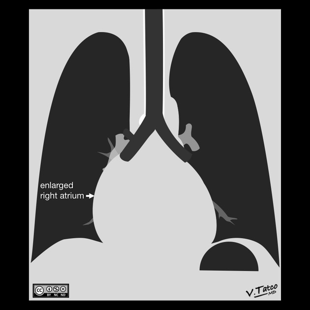
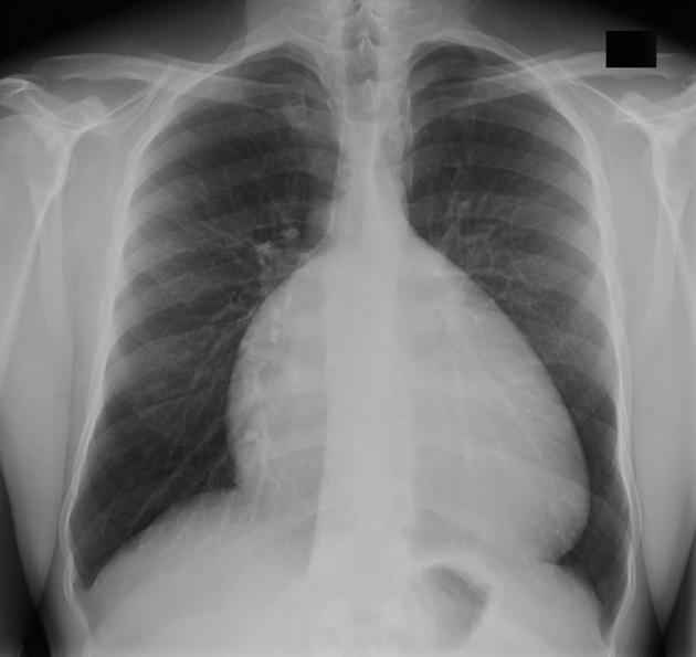
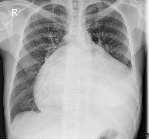
On a frontal view, the right atrium is visible because of its interface with the right middle lobe. Subtle and moderate right atrial enlargement is not accurately determined on plain films because there is normal variability in the shape of the right atrium. Features are non-specific but include 2,3:
enlarged, globular heart
narrow vascular pedicle
-
gross enlargement of the right atrial shadow i.e. increased convexity in the lower half of the right cardiac border
right atrial convexity is more than 50% of the cardiovascular height
right atrial margin is more than 5.5 cm from the midline
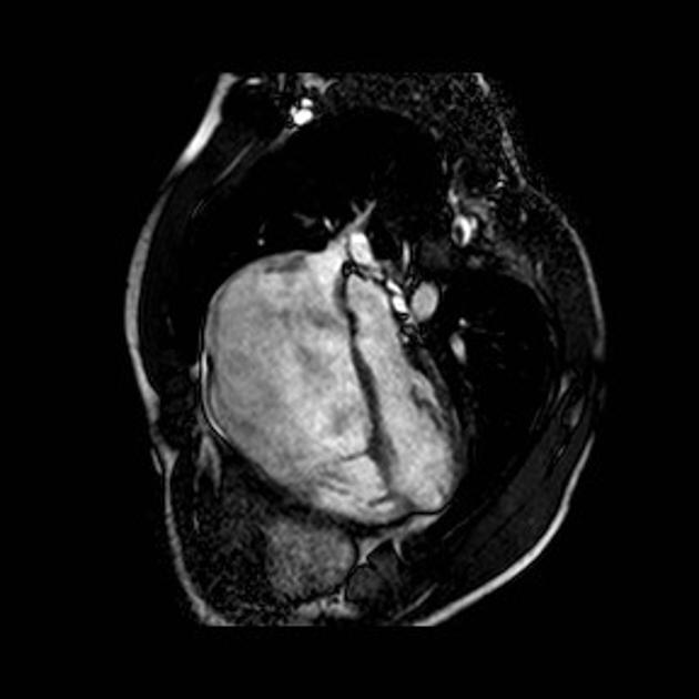
CT/MRI
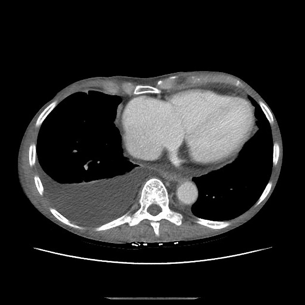
There are no accepted standard reference measurements, and enlargement is usually determined qualitatively on CT or MRI. Right atrial volume is not routinely recorded on echocardiography. Suggested measurements include 5:
-
right atrial normal size (measured at end-systole on four-chamber view)
long axis: 3.4-5.3 cm
short axis: 2.6-4.4 cm
-
area: 10-18 cm2
echocardiography estimates tend to be larger than on CT or MRI
Ultrasound
Echocardiography
With transthoracic echocardiography, the apical 4-chamber view is preferred to assess right atrial (RA) dimensions by tracing the chamber margins at end-systole and recording orthogonal measurements of the major and minor axes. These dimensions may be acquired as follows 6:
-
major (long) axis
taken parallel to the interatrial septum from the center of the tricuspid annular plane to the central superior wall of the RA
mean RA major axis is 4.4 cm, considered enlarged when >5.3 cm
-
minor (short) axis
measured at the mid right atrium perpendicular to the long axis from the free wall to the interatrial septum
mean RA minor axis is 3.5 cm, considered enlarged when >4.5 cm
-
area
tracing should encompass the tricuspid annular plane interatrial septum, and the endocardial borders of the superior and free right atrial walls
care should be taken to avoid inclusion of the tricuspid valve leaflets and annulus, the right atrial appendage, as well as the superior and inferior vena cava
mean RA area is 14 cm2, considered enlarged when >18 cm2


 Unable to process the form. Check for errors and try again.