
Congenital urachal anomalies
Citation, DOI, disclosures and article data
At the time the article was created Yuranga Weerakkody had no recorded disclosures.
View Yuranga Weerakkody's current disclosuresAt the time the article was last revised Mohammad Taghi Niknejad had no financial relationships to ineligible companies to disclose.
View Mohammad Taghi Niknejad's current disclosures- Congenital urachal remnant anomalies
- Urachal remnant abnormalities
- Urachal remnant anomalies
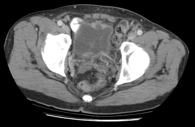
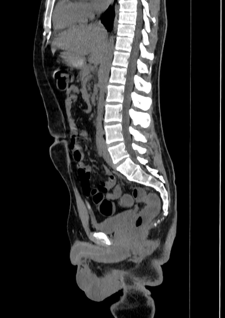
Congenital urachal anomalies are a spectrum of potential anomalies that can occur due to incomplete involution of the urachus.
On this page:
Epidemiology
A urachal remnant occurs in approximately 1 in 5000 patients.
Pathology
The urachus connects the dome of the bladder to the umbilical cord during fetal life and is located behind the lower anterior abdominal wall and anterior to the peritoneum in the space of Retzius.
By birth, the urachus is obliterated and becomes a vestigial structure known as the median umbilical ligament (not to be confused with the medial umbilical ligament, which is a separate structure that lies laterally to the median umbilical ligament).
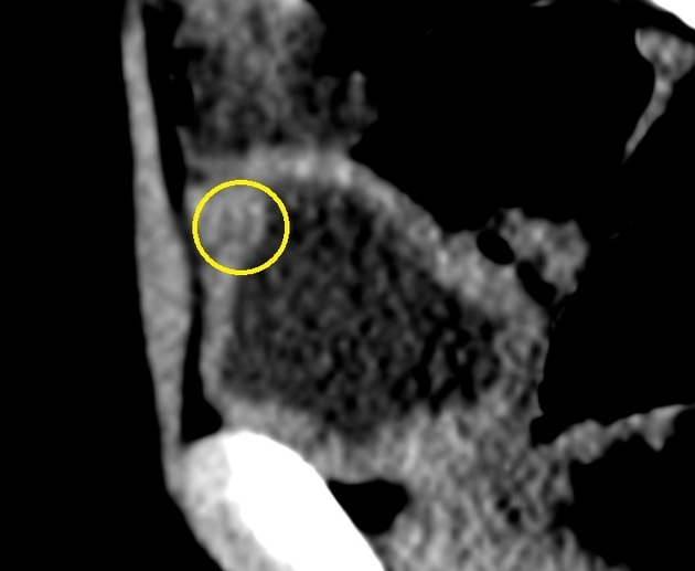
In the absence of complete obliteration, the urachus persists in a number of configurations depending on the location and degree of obliteration.
Types
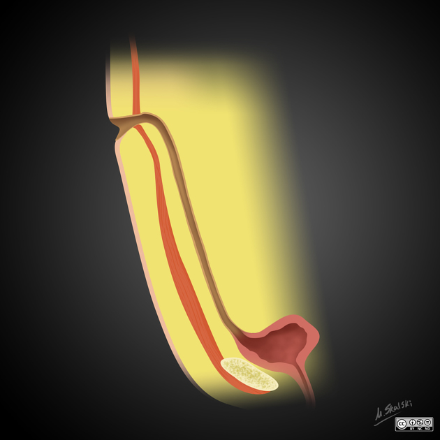
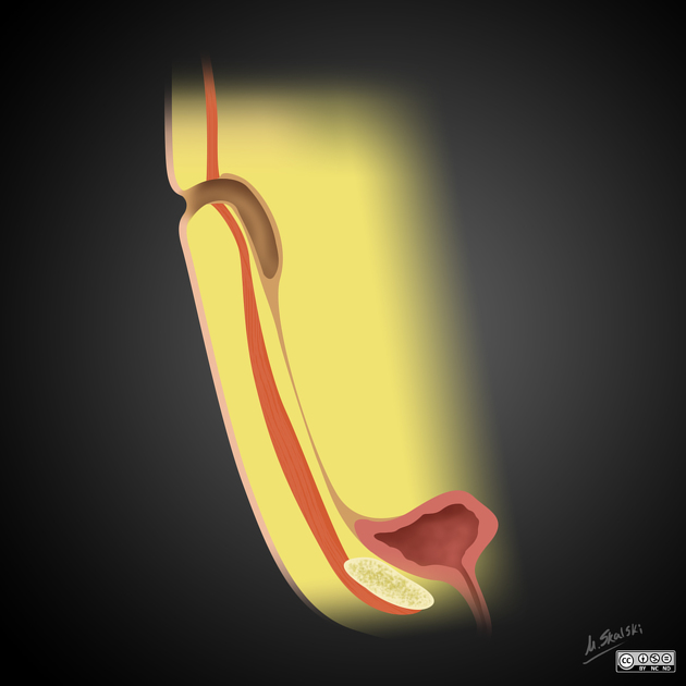
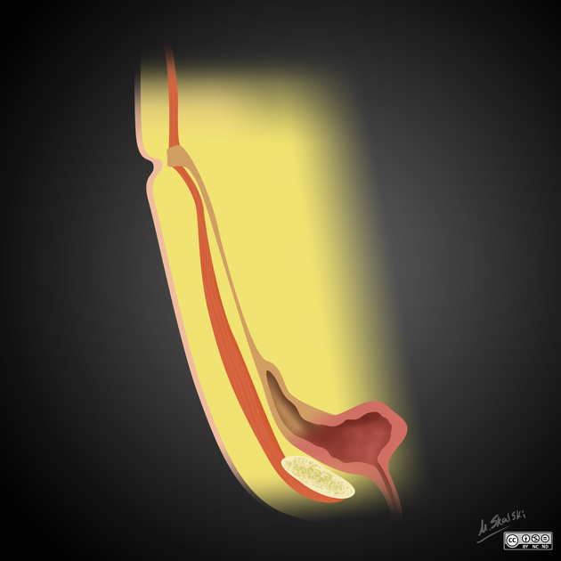
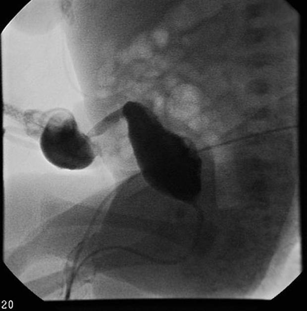
There are four types of congenital urachal remnant anomalies. They are:
-
communication between the bladder and umbilicus through a urachus that has not involuted
commonest (~50%)
-
a fluid-filled dilatation of the mid urachus
next commonest (~30%)
-
blind focal dilatation of the umbilical end of the urachus
~15%
-
blind focal dilatation of the bladder end of the urachus
~5%
Treatment and prognosis
Congenital urachal remnants predispose to infection from urinary stasis and over a long period the remnant may develop malignancy (e.g. adenocarcinoma, sarcoma 6,7).
Treatment is not standardized. Some recommend surgical excision of the urachus if a remnant anomaly is present.
References
- 1. Parada Villavicencio C, Adam SZ, Nikolaidis P, Yaghmai V, Miller FH. Imaging of the Urachus: Anomalies, Complications, and Mimics. Radiographics : a review publication of the Radiological Society of North America, Inc. 36 (7): 2049-2063. doi:10.1148/rg.2016160062 - Pubmed
- 2. Yu JS, Kim KW, Lee HJ et-al. Urachal remnant diseases: spectrum of CT and US findings. Radiographics. 21 (2): 451-61. Radiographics (full text) - Pubmed citation
- 3. Donnelly LF, Frush DP. Cross-sectional imaging of abnormalities of the abdominal wall in pediatric patients. AJR Am J Roentgenol. 2001;176 (5): 1233-9. AJR Am J Roentgenol (full text) - Pubmed citation
- 4. Disantis DJ, Siegel MJ, Katz ME. Simplified approach to umbilical remnant abnormalities. Radiographics. 1991;11 (1): 59-66. Radiographics (abstract) - Pubmed citation
- 5. Robert Y, Hennequin-Delerue C, Chaillet D, Dubrulle F, Biserte J, Lemaitre L. Urachal remnants: sonographic assessment. (1996) Journal of clinical ultrasound : JCU. 24 (7): 339-44. doi:10.1002/(SICI)1097-0096(199609)24:7<339::AID-JCU2>3.0.CO;2-C - Pubmed
- 6. Karray A, Sahli S, Rahal Z, Aziza B, Jouini R. Urachal Rhabdomyosarcoma: A Case Report of an Extremely Rare Localisation. Urol Case Rep. 2022;43:102109. doi:10.1016/j.eucr.2022.102109 - Pubmed
- 7. Rhudd A, Moghul M, Nair G, McDonald J. Malignant Transformation of a Urachal Cyst—a Case Report and Literature Review. Journal of Surgical Case Reports. 2018;2018(3):rjy056. doi:10.1093/jscr/rjy056 - Pubmed
Incoming Links
- Diffuse adenomyosis
- Infected urachal cyst with sinus formation
- Vesicourachal diverticulum
- Vesicourachal diverticulum and calculus
- Vesicourachal diverticulum
- Infected urachal cyst with urachal-sigmoid fistula
- Urachal cyst
- Patent urachus (ultrasound)
- Patent urachus (ultrasound)
- Urachal carcinoma
- Urachal carcinoma
- Urachus (illustration)
- Infected vesicourachal diverticulum
- Complicated urachal remnant
- Infected urachal cyst
- Infected patent urachus
- Median umbilical ligament
- Vesicourachal diverticulum
Related articles: Pathology: Genitourinary
- obstetrics
-
first trimester
- ultrasound findings in early pregnancy
- embryo/fetus
- beta-hCG levels
- confirming intrauterine gestation
- pregnancy of unknown location (PUL)
- first trimester vaginal bleeding
- early structural scan
- aneuploidy testing
-
second trimester
- fetal biometry
- amniotic fluid volume
- fetal morphology assessment
- soft markers
- amnioreduction
- Doppler ultrasound
- nuchal translucency
- 11-13 weeks antenatal scan
- chorionic villus sampling (CVS) and amniocentesis
- other
- placenta
- placental anatomy
- placental developmental abnormalities
- placenta previa
- spectrum of abnormal placental villous adherence
- abnormalities of cord insertion
- abruptio placentae
- placental pathology
- vascular pathologies of placenta
- placental infections
- placental masses
- molar pregnancy
- twin placenta
- miscellaneous
-
first trimester
- gynecology
- acute pelvic pain
- chronic pelvic pain
- uterus
- ovaries
- ovarian follicle
- ovarian torsion
- pelvic inflammatory disease
- ovarian cysts and masses
- paraovarian cyst
- polycystic ovaries
- ovarian hyperstimulation syndrome
- post-hysterectomy ovary
- cervix
- fallopian tube
- other
- male genital tract
- prostate gland
- transrectal ultrasound
- prostate tumors
- infections of the prostate
-
prostatitis
- acute bacterial prostatitis
-
chronic prostatitis
- chronic bacterial prostatitis
- chronic prostatitis and chronic pelvic pain syndrome (CPPS)
- asymptomatic inflammatory prostatitis
- granulomatous prostatitis
- emphysematous prostatitis
- prostatic abscess
-
prostatitis
- benign prostatic hypertrophy
- cystic lesions of the prostate
- prostatic calcification
- prostatic infarction
- testes
-
unilateral testicular lesion
- testicular torsion
- orchitis
- testicular trauma
-
germ cell tumors of the testis
- testicular seminoma
-
non seminomatous germ cell tumors
- mixed germ cell tumor
- yolk sac tumor (endodermal sinus tumor)
- embryonal cell carcinoma
- choriocarcinoma
- testicular teratoma
- testicular epidermoid (teratoma with ectodermal elements only)
- burned out testis tumor
- sex cord / stromal tumors of the testis
- testicular cyst
- testicular lymphoma
- bilateral testicular lesion
- paratesticular lesions
- epididymis
- other
- polyorchidism
- cryptorchidism
- tubular ectasia of the rete testis
- cystadenoma of the rete testis
- testicular sarcoidosis
- testicular tuberculosis
- spermatic cord
- fibrous pseudotumor of the scrotum
- scrotal leiomyosarcoma
- testicular adrenal rest tumors (TARTs)
- tunica vaginalis testis mesothelioma
- splenogonadal fusion
- testicular vasculitis
- abnormal testicular Doppler flow (differential)
-
unilateral testicular lesion
- penis
- prostate gland
- KUB
- kidneys
- normal renal anatomy
- hydronephrosis
- urolithiasis
- renal masses
- renal cystic disease
- renal infection
- vascular
- trauma
- ureter
- normal ureter anatomy
- ureteral stricture
- ureteral dilatation
- ureteral anomalies
- ureteral tumors
- ureteral trauma
- other
- bladder
- kidneys


 Unable to process the form. Check for errors and try again.