
Moiré fringes
Citation, DOI, disclosures and article data
At the time the article was created J. Ray Ballinger had no recorded disclosures.
View J. Ray Ballinger's current disclosuresAt the time the article was last revised Candace Makeda Moore had no financial relationships to ineligible companies to disclose.
View Candace Makeda Moore's current disclosures- Moire fringes
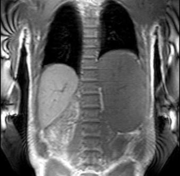
Moiré fringes are an interference pattern most commonly seen when acquiring gradient echo images using the body coil.
Because of the lack of perfect homogeneity of the main magnetic field from one side of the body to the other, aliasing of one side of the body to the other results in the superimposition of signals of different phases that alternatively add and cancel 1,2, this causes the banding appearance similar to the effect of looking through two screen windows or through the railings of bridge from distance.
Shimming will help to reduce this artifact by making the magnetic field more homogeneous 4.
The term Moiré when used in digital imaging and computer graphics describes an artefact that can be created by overlaying two semi-transparent grids or repeating line patterns on each other, which creates an interference pattern. In general radiography, the term has been used to describe the 'grid moiré pattern' where there is under sampling due to incorrect grid placement or alignment 3.
References
- 1. Stadler A, Schima W, Ba-Ssalamah A, Kettenbach J, Eisenhuber E. Artifacts in Body MR Imaging: Their Appearance and How to Eliminate Them. Eur Radiol. 2006;17(5):1242-55. doi:10.1007/s00330-006-0470-4 - Pubmed
- 2. Ho C, Xiao L, Kwok K et al. Common Artifacts in Magnetic Resonance Imaging: A Pictorial Essay. Hong Kong Journal of Radiology. 2023;26(1):58-65. doi:10.12809/hkjr2317476
- 3. Lin C, Lee W, Chen S et al. A Study of Grid Artifacts Formation and Elimination in Computed Radiographic Images. J Digit Imaging. 2006;19(4):351-61. doi:10.1007/s10278-006-0630-8 - Pubmed
- 4. Wachowicz, K. (2014). Evaluation of active and passive shimming in magnetic resonance imaging. Research and Reports in Nuclear Medicine, 1.
Incoming Links
Related articles: Imaging technology
- imaging technology
- imaging physics
- imaging in practice
-
x-rays
- x-ray physics
- x-ray in practice
- x-ray production
- x-ray tube
- filters
- automatic exposure control (AEC)
- beam collimators
- grids
- air gap technique
- cassette
- intensifying screen
- x-ray film
- image intensifier
- digital radiography
- digital image
- mammography
- x-ray artifacts
- radiation units
- radiation safety
- radiation detectors
- fluoroscopy
-
computed tomography (CT)
- CT physics
- CT in practice
- CT technology
- CT image reconstruction
- CT image quality
- CT dose
-
CT contrast media
-
iodinated contrast media
- agents
- water soluble
- water insoluble
- vicarious contrast material excretion
- iodinated contrast media adverse reactions
- agents
- non-iodinated contrast media
-
iodinated contrast media
-
CT artifacts
- patient-based artifacts
- physics-based artifacts
- hardware-based artifacts
- ring artifact
- tube arcing
- out of field artifact
- air bubble artifact
- helical and multichannel artifacts
- CT safety
- history of CT
-
MRI
- MRI physics
- MRI in practice
- MRI hardware
- signal processing
-
MRI pulse sequences (basics | abbreviations | parameters)
- T1 weighted image
- T2 weighted image
- proton density weighted image
- chemical exchange saturation transfer
- CSF flow studies
- diffusion weighted imaging (DWI)
- echo-planar pulse sequences
- fat-suppressed imaging sequences
- gradient echo sequences
- inversion recovery sequences
- metal artifact reduction sequence (MARS)
-
perfusion-weighted imaging
- techniques
- derived values
- saturation recovery sequences
- spin echo sequences
- spiral pulse sequences
- susceptibility-weighted imaging (SWI)
- T1 rho
- MR angiography (and venography)
-
MR spectroscopy (MRS)
- 2-hydroxyglutarate peak: resonates at 2.25 ppm
- alanine peak: resonates at 1.48 ppm
- choline peak: resonates at 3.2 ppm
- citrate peak: resonates at 2.6 ppm
- creatine peak: resonates at 3.0 ppm
- functional MRI (fMRI)
- gamma-aminobutyric acid (GABA) peak: resonates at 2.2-2.4 ppm
- glutamine-glutamate peak: resonates at 2.2-2.4 ppm
- Hunter's angle
- lactate peak: resonates at 1.3 ppm
- lipids peak: resonates at 1.3 ppm
- myoinositol peak: resonates at 3.5 ppm
- MR fingerprinting
- N-acetylaspartate (NAA) peak: resonates at 2.0 ppm
- propylene glycol peak: resonates at 1.13 ppm
-
MRI artifacts
- MRI hardware and room shielding
- MRI software
- patient and physiologic motion
- tissue heterogeneity and foreign bodies
- Fourier transform and Nyquist sampling theorem
- MRI contrast agents
- MRI safety
-
ultrasound
- ultrasound physics
-
transducers
- linear array
- convex array
- phased array
- frame averaging (frame persistence)
- ultrasound image resolution
- imaging modes and display
- pulse-echo imaging
- real-time imaging
-
Doppler imaging
- Doppler effect
- color Doppler
- power Doppler
- B flow
- color box
- Doppler angle
- pulse repetition frequency and scale
- wall filter
- color write priority
- packet size (dwell time)
- peak systolic velocity
- end-diastolic velocity
- resistive index
- pulsatility index
- Reynolds number
- panoramic imaging
- compound imaging
- harmonic imaging
- elastography
- scanning modes
- 2D ultrasound
- 3D ultrasound
- 4D ultrasound
- M-mode
-
ultrasound artifacts
- acoustic shadowing
- acoustic enhancement
- beam width artifact
- reverberation artifact
- ring down artifact
- mirror image artifact
- side lobe artifact
- speckle artifact
- speed displacement artifact
- refraction artifact
- multipath artifact
- anisotropy
- electrical interference artifact
- hardware-related artifacts
- Doppler artifacts
- aliasing
- tissue vibration
- spectral broadening
- blooming
- motion (flash) artifact
- twinkling artifact
- acoustic streaming
- biological effects of ultrasound
- history of ultrasound
-
nuclear medicine
- nuclear medicine physics
- detectors
- tissue to background ratio
-
radiopharmaceuticals
- fundamentals of radiopharmaceuticals
- radiopharmaceutical labeling
- radiopharmaceutical production
- nuclear reactor produced radionuclides
- cyclotron produced radionuclides
- radiation detection
- dosimetry
- specific agents
- carbon-11
- chromium-51
- fluorine agents
- gallium agents
- Ga-67 citrate
- Ga-68
- iodine agents
-
I-123
- I-123 iodide
- I-123 ioflupane (DaTSCAN)
- I-123 ortho-iodohippurate
- I-131
-
MIBG scans
- I-123 MIBG
- I-131 MIBG
-
I-123
- indium agents
- In-111 Octreoscan
- In-111 OncoScint
- In-111 Prostascint
- In-111 oxine labeled WBC
- krypton-81m
- nitrogen-13
- oxygen-15
- phosphorus-32
- selenium-75
-
technetium agents
- Tc-99m DMSA
- Tc-99m DTPA
- Tc-99m DTPA aerosol
- Tc-99m HMPAO
- Tc-99m HMPAO labeled WBC
- Tc-99m MAA
- Tc-99m MAG3
- Tc-99m MDP
- Tc-99m mercaptoacetyltriglycine
- Tc-99m pertechnetate
- Tc-99m labeled RBC
- Tc-99m sestamibi
- Tc-99m sulfur colloid
- Tc-99m sulfur colloid (oral)
- thallium-201 chloride
- xenon agents
- in vivo therapeutic agents
- pharmaceuticals used in nuclear medicine
-
emerging methods in medical imaging
- radiography
- phase-contrast imaging
- CT
- deep-learning reconstruction
- photon counting CT
- virtual non-contrast imaging
- ultrasound
- magnetomotive ultrasound (MMUS)
- superb microvascular imaging
- ultrafast Doppler imaging
- ultrasound localization microscopy
- MRI
- nuclear medicine
- total body PET system
- immuno-PET
- miscellaneous
- radiography


 Unable to process the form. Check for errors and try again.