
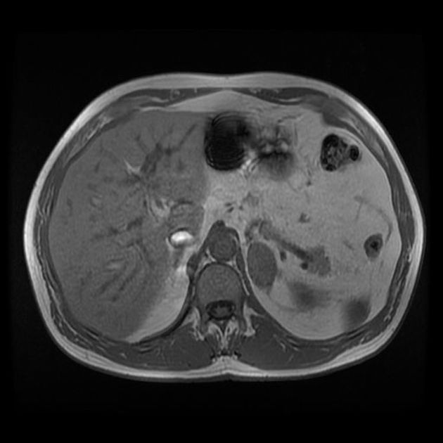
T1 weighted image
Citation, DOI, disclosures and article data
At the time the article was created Jeremy Jones had no recorded disclosures.
View Jeremy Jones's current disclosuresAt the time the article was last revised Tariq Walizai had no financial relationships to ineligible companies to disclose.
View Tariq Walizai's current disclosures- T1WI
- T1
- Spin-lattice
- T1-weighted image
- T1W imaging
- T1 weighted images
T1 weighted image (also referred to as T1WI or the "spin-lattice" relaxation time) is one of the basic pulse sequences in MRI and demonstrates differences in the T1 relaxation times of tissues.
A T1WI relies upon the longitudinal relaxation of a tissue's net magnetization vector (NMV). Basically, spins aligned in an external field (B0) are put into the transverse plane by a radiofrequency (RF) pulse. They then slide back toward the original equilibrium of B0. Not all tissues return back to equilibrium in the same amount of time, and a tissue's T1 reflects the amount of time taken for its protons' spins to realign with the main magnetic field (B0).
T1 weighting tends to have short TE and TR times.
Fat quickly realigns its longitudinal magnetization with B0, and it therefore appears bright on a T1 weighted image. Conversely, water has much slower longitudinal magnetization realignment after an RF pulse and therefore, has less transverse magnetization after an RF pulse. Thus, water has low signal and appears dark.
If T1WIs did not have short TRs, then all the protons would recover their alignment with the main magnetic field and the image would be uniformly intense. Selecting a TR shorter than the tissues' recovery time allows one to differentiate them (i.e. tissue contrast).
T1-weighted sequences provide the best contrast for paramagnetic contrast agents (e.g. gadolinium-containing compounds).
T1-weighted sequences include:
T1W spin echo (SE)
T1W gradient echo (GRE)
gadolinium postcontrast sequences (gradient echo sequences)
time of flight 2D or 3D MR angiography sequences
ADVERTISEMENT: Supporters see fewer/no ads
Practical tips
-
signal hyperintensity on T1WI is an important finding and needs to be explained, the potential causes of this appearance are:
fat
paramagnetic contrast media e.g. gadolinium-based agents
melanin
slow-flowing blood
proteinaceous fluid
calcium 3
copper 3
manganese 3
iron 3
Summary
TR: short
TE: short
fat: bright
fluid: dark
References
- 1. Mitchell DG, Cohen M. MRI principles. Saunders. ISBN:0721600247. Read it at Google Books - Find it at Amazon
- 2. Westbrook C. MRI at a glance. Wiley-Blackwell. ISBN:0632056193. Read it at Google Books - Find it at Amazon
- 3. Kinoshita T, Ogawa T, Yoshida Y, Tamura H, Kado H, Okudera T. Curvilinear T1 hyperintense lesions representing cortical necrosis after cerebral infarction. Neuroradiology. 2005 Sep;47(9):647-51. doi: 10.1007/s00234-005-1398-0. Epub 2005 Jul 19. PMID: 16028037
Incoming Links
- 3D fast spin echo (MRI sequence)
- Thoracic spine protocol (MRI)
- Nuclear magnetisation
- Hyperintense on T1-weighted images (mnemonic)
- Shoulder protocol (MRI)
- Flip-flop effect
- MR enterography
- White matter tracts
- T1 mapping - myocardium
- Oncocytic sinonasal papilloma
- Bright tongue sign
- Lumbar spine protocol (MRI)
- Mid- and forefoot protocol (MRI)
- T1 black holes
- Elbow protocol (MRI)
- T1 relaxation time
- Late gadolinium enhancement
- Hip protocol (MRI)
- Cartilage injury (overview)
- Relaxation
Related articles: Imaging technology
- imaging technology
- imaging physics
- imaging in practice
-
x-rays
- x-ray physics
- x-ray in practice
- x-ray production
- x-ray tube
- filters
- automatic exposure control (AEC)
- beam collimators
- grids
- air gap technique
- cassette
- intensifying screen
- x-ray film
- image intensifier
- digital radiography
- digital image
- mammography
- x-ray artifacts
- radiation units
- radiation safety
- radiation detectors
- fluoroscopy
-
computed tomography (CT)
- CT physics
- CT in practice
- CT technology
- CT image reconstruction
- CT image quality
- CT dose
-
CT contrast media
-
iodinated contrast media
- agents
- water soluble
- water insoluble
- vicarious contrast material excretion
- iodinated contrast media adverse reactions
- agents
- non-iodinated contrast media
-
iodinated contrast media
-
CT artifacts
- patient-based artifacts
- physics-based artifacts
- hardware-based artifacts
- ring artifact
- tube arcing
- out of field artifact
- air bubble artifact
- helical and multichannel artifacts
- CT safety
- history of CT
-
MRI
- MRI physics
- MRI in practice
- MRI hardware
- signal processing
-
MRI pulse sequences (basics | abbreviations | parameters)
- T1 weighted image
- T2 weighted image
- proton density weighted image
- chemical exchange saturation transfer
- CSF flow studies
- diffusion weighted imaging (DWI)
- echo-planar pulse sequences
- fat-suppressed imaging sequences
- gradient echo sequences
- inversion recovery sequences
- metal artifact reduction sequence (MARS)
-
perfusion-weighted imaging
- techniques
- derived values
- saturation recovery sequences
- spin echo sequences
- spiral pulse sequences
- susceptibility-weighted imaging (SWI)
- T1 rho
- MR angiography (and venography)
-
MR spectroscopy (MRS)
- 2-hydroxyglutarate peak: resonates at 2.25 ppm
- alanine peak: resonates at 1.48 ppm
- choline peak: resonates at 3.2 ppm
- citrate peak: resonates at 2.6 ppm
- creatine peak: resonates at 3.0 ppm
- functional MRI (fMRI)
- gamma-aminobutyric acid (GABA) peak: resonates at 2.2-2.4 ppm
- glutamine-glutamate peak: resonates at 2.2-2.4 ppm
- Hunter's angle
- lactate peak: resonates at 1.3 ppm
- lipids peak: resonates at 1.3 ppm
- myoinositol peak: resonates at 3.5 ppm
- MR fingerprinting
- N-acetylaspartate (NAA) peak: resonates at 2.0 ppm
- propylene glycol peak: resonates at 1.13 ppm
-
MRI artifacts
- MRI hardware and room shielding
- MRI software
- patient and physiologic motion
- tissue heterogeneity and foreign bodies
- Fourier transform and Nyquist sampling theorem
- MRI contrast agents
- MRI safety
-
ultrasound
- ultrasound physics
-
transducers
- linear array
- convex array
- phased array
- frame averaging (frame persistence)
- ultrasound image resolution
- imaging modes and display
- pulse-echo imaging
- real-time imaging
-
Doppler imaging
- Doppler effect
- color Doppler
- power Doppler
- B flow
- color box
- Doppler angle
- pulse repetition frequency and scale
- wall filter
- color write priority
- packet size (dwell time)
- peak systolic velocity
- end-diastolic velocity
- resistive index
- pulsatility index
- Reynolds number
- panoramic imaging
- compound imaging
- harmonic imaging
- elastography
- scanning modes
- 2D ultrasound
- 3D ultrasound
- 4D ultrasound
- M-mode
-
ultrasound artifacts
- acoustic shadowing
- acoustic enhancement
- beam width artifact
- reverberation artifact
- ring down artifact
- mirror image artifact
- side lobe artifact
- speckle artifact
- speed displacement artifact
- refraction artifact
- multipath artifact
- anisotropy
- electrical interference artifact
- hardware-related artifacts
- Doppler artifacts
- aliasing
- tissue vibration
- spectral broadening
- blooming
- motion (flash) artifact
- twinkling artifact
- acoustic streaming
- biological effects of ultrasound
- history of ultrasound
-
nuclear medicine
- nuclear medicine physics
- detectors
- tissue to background ratio
-
radiopharmaceuticals
- fundamentals of radiopharmaceuticals
- radiopharmaceutical labeling
- radiopharmaceutical production
- nuclear reactor produced radionuclides
- cyclotron produced radionuclides
- radiation detection
- dosimetry
- specific agents
- carbon-11
- chromium-51
- fluorine agents
- gallium agents
- Ga-67 citrate
- Ga-68
- iodine agents
-
I-123
- I-123 iodide
- I-123 ioflupane (DaTSCAN)
- I-123 ortho-iodohippurate
- I-131
-
MIBG scans
- I-123 MIBG
- I-131 MIBG
-
I-123
- indium agents
- In-111 Octreoscan
- In-111 OncoScint
- In-111 Prostascint
- In-111 oxine labeled WBC
- krypton-81m
- nitrogen-13
- oxygen-15
- phosphorus-32
- selenium-75
-
technetium agents
- Tc-99m DMSA
- Tc-99m DTPA
- Tc-99m DTPA aerosol
- Tc-99m HMPAO
- Tc-99m HMPAO labeled WBC
- Tc-99m MAA
- Tc-99m MAG3
- Tc-99m MDP
- Tc-99m mercaptoacetyltriglycine
- Tc-99m pertechnetate
- Tc-99m labeled RBC
- Tc-99m sestamibi
- Tc-99m sulfur colloid
- Tc-99m sulfur colloid (oral)
- thallium-201 chloride
- xenon agents
- in vivo therapeutic agents
- pharmaceuticals used in nuclear medicine
-
emerging methods in medical imaging
- radiography
- phase-contrast imaging
- CT
- deep-learning reconstruction
- photon counting CT
- virtual non-contrast imaging
- ultrasound
- magnetomotive ultrasound (MMUS)
- superb microvascular imaging
- ultrafast Doppler imaging
- ultrasound localization microscopy
- MRI
- nuclear medicine
- total body PET system
- immuno-PET
- miscellaneous
- radiography


 Unable to process the form. Check for errors and try again.