
Echogenic intracardiac focus
Citation, DOI, disclosures and article data
At the time the article was created Yuranga Weerakkody had no recorded disclosures.
View Yuranga Weerakkody's current disclosuresAt the time the article was last revised Arlene Campos had no financial relationships to ineligible companies to disclose.
View Arlene Campos's current disclosures- Echogenic intracardiac focus (EIF)
- Echogenic cardiac focus
- Intracardiac echogenic focus
- Intracardiac echogenic focus (IEF)
- Echogenic focus in intrapapillary muscle
- Echogenic intracardiac foci
- Echogenic intra-cardiac focus
- Echogenic intra-cardiac foci
Echogenic intracardiac focus (EIF) is a relatively common sonographic observation that may be present on an antenatal ultrasound scan.
On this page:
Epidemiology
They are thought to be present in ~4-5% of karyotypically normal fetuses. They may be more common in the Asian population 5.
Associations
trisomy 21 (Down syndrome): may be present in up to 12% of fetuses
The tightness of the association between an isolated echogenic intracardiac focus and aneuploidy continues to be debated. Biventricular echogenic intracardiac foci are considered to be a higher risk for aneuploidy.
Pathology
They are considered to represent mineralization within the papillary muscles.
Location
The majority of echogenic intracardiac foci are unilateral. Out of all the cardiac chambers, the left ventricle is the most frequent in terms of location.
Radiographic features
Ultrasound
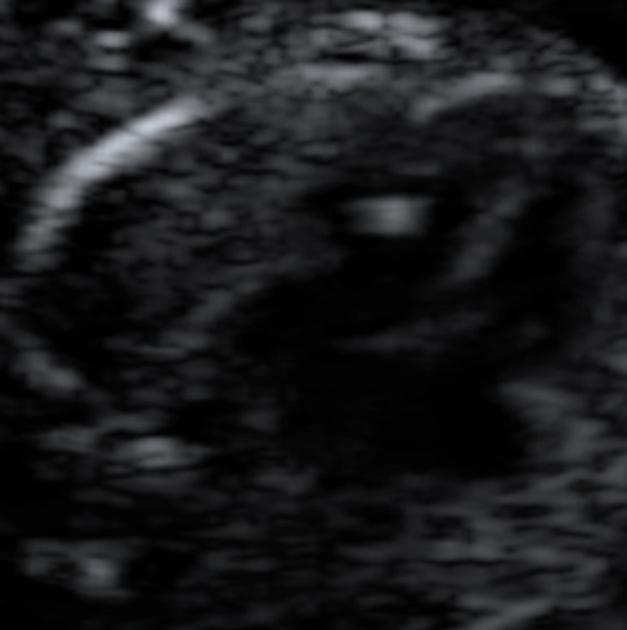
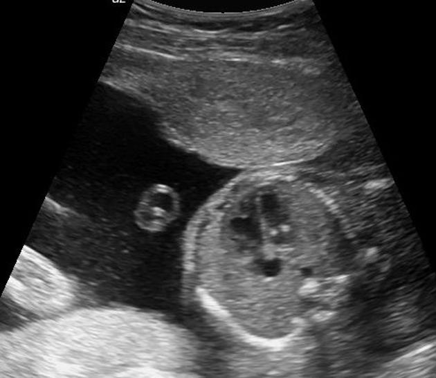
They are typically seen as a small bright echogenic focus within the fetal heart on a four chamber view (often as bright as bone).
Tissue harmonic imaging should be turned off when evaluating a potential echogenic intracardiac focus, to avoid false positives. If it is difficult to tell if the focus is as bright as bone, the gain in the image can be decreased to see which structure disappears first.
It is usually single and <3 mm.
It needs to be differentiated from normal papillary muscle which is not as bright as bone and a moderator band which is situated at the ventricular apex.
ADVERTISEMENT: Supporters see fewer/no ads
Treatment and prognosis
The presence of an echogenic intracardiac focus has to be interpreted in the context of maternal risk factors and other sonographic anomalies.
When seen incidentally in isolation in a normal pregnancy, it is considered a benign variant and some authors state karyotyping is unwarranted in the mid-trimester 8. There is no recognized direct association with congenital heart anomalies for an isolated echogenic intracardiac focus 1 (unless there is an associated aneuploidic anomaly).
In high-risk pregnancies, there is an increased incidence of aneuploidy (e.g. Down syndrome 2,3 and trisomy 13 3) and it is classified as a soft marker. The presence of multiple or bilateral (more than one chamber) echogenic foci is considered to increase the risk.
They usually disappear during the 3rd trimester 7.
References
- 1. Wax J, Cartin A, Pinette M, Blackstone J. Are Intracardiac Echogenic Foci Markers of Congenital Heart Disease in the Fetus with Chromosomal Abnormalities? J Ultrasound Med. 2004;23(7):895-8. doi:10.7863/jum.2004.23.7.895 - Pubmed
- 2. Manning J, Ragavendra N, Sayre J et al. Significance of Fetal Intracardiac Echogenic Foci in Relation to Trisomy 21: A Prospective Sonographic Study of High-Risk Pregnant Women. AJR Am J Roentgenol. 1998;170(4):1083-4. doi:10.2214/ajr.170.4.9530064 - Pubmed
- 3. Winter TC, Anderson AM, Cheng EY et-al. Echogenic intracardiac focus in 2nd-trimester fetuses with trisomy 21: usefulness as a US marker. Radiology. 2000;216 (2): 450-6. Radiology (full text) - Pubmed citation
- 4. Bradley K, Santulli T, Gregory K, Herbert W, Carlson D, Platt L. An Isolated Intracardiac Echogenic Focus as a Marker for Aneuploidy. Am J Obstet Gynecol. 2005;192(6):2021-6; discussion 2026. doi:10.1016/j.ajog.2005.03.033 - Pubmed
- 5. Shipp TD, Bromley B, Lieberman E et-al. The frequency of the detection of fetal echogenic intracardiac foci with respect to maternal race. Ultrasound Obstet Gynecol. 2000;15 (6): 460-2. doi:10.1046/j.1469-0705.2000.00138.x - Pubmed citation
- 6. Carriço A, Matias A, Areias J. How Important is a Cardiac Echogenic Focus in a Routine Fetal Examination? Rev Port Cardiol. 2004;23(3):459-61. - Pubmed
- 7. Michael Entezami. Ultrasound Diagnosis of Fetal Anomalies. (2004) ISBN: 9781588902122 - Google Books
- 8. Achiron R, Lipitz S, Gabbay U, Yagel S. Prenatal Ultrasonographic Diagnosis of Fetal Heart Echogenic Foci: No Correlation with Down Syndrome. Obstet Gynecol. 1997;89(6):945-8. doi:10.1016/s0029-7844(97)00131-2 - Pubmed
- 9. Anderson N & Jyoti R. Relationship of Isolated Fetal Intracardiac Echogenic Focus to Trisomy 21 at the Mid-Trimester Sonogram in Women Younger Than 35 Years. Ultrasound Obstet Gynecol. 2003;21(4):354-8. doi:10.1002/uog.89 - Pubmed
Incoming Links
Related articles: Pathology: Genitourinary
- obstetrics
-
first trimester
- ultrasound findings in early pregnancy
- embryo/fetus
- beta-hCG levels
- confirming intrauterine gestation
- pregnancy of unknown location (PUL)
- first trimester vaginal bleeding
- early structural scan
- aneuploidy testing
-
second trimester
- fetal biometry
- amniotic fluid volume
- fetal morphology assessment
- soft markers
- amnioreduction
- Doppler ultrasound
- nuchal translucency
- 11-13 weeks antenatal scan
- chorionic villus sampling (CVS) and amniocentesis
- other
- placenta
- placental anatomy
- placental developmental abnormalities
- placenta previa
- spectrum of abnormal placental villous adherence
- abnormalities of cord insertion
- abruptio placentae
- placental pathology
- vascular pathologies of placenta
- placental infections
- placental masses
- molar pregnancy
- twin placenta
- miscellaneous
-
first trimester
- gynecology
- acute pelvic pain
- chronic pelvic pain
- uterus
- ovaries
- ovarian follicle
- ovarian torsion
- pelvic inflammatory disease
- ovarian cysts and masses
- paraovarian cyst
- polycystic ovaries
- ovarian hyperstimulation syndrome
- post-hysterectomy ovary
- cervix
- fallopian tube
- other
- male genital tract
- prostate gland
- transrectal ultrasound
- prostate tumors
- infections of the prostate
-
prostatitis
- acute bacterial prostatitis
-
chronic prostatitis
- chronic bacterial prostatitis
- chronic prostatitis and chronic pelvic pain syndrome (CPPS)
- asymptomatic inflammatory prostatitis
- granulomatous prostatitis
- emphysematous prostatitis
- prostatic abscess
-
prostatitis
- benign prostatic hypertrophy
- cystic lesions of the prostate
- prostatic calcification
- prostatic infarction
- testes
-
unilateral testicular lesion
- testicular torsion
- orchitis
- testicular trauma
-
germ cell tumors of the testis
- testicular seminoma
-
non seminomatous germ cell tumors
- mixed germ cell tumor
- yolk sac tumor (endodermal sinus tumor)
- embryonal cell carcinoma
- choriocarcinoma
- testicular teratoma
- testicular epidermoid (teratoma with ectodermal elements only)
- burned out testis tumor
- sex cord / stromal tumors of the testis
- testicular cyst
- testicular lymphoma
- bilateral testicular lesion
- paratesticular lesions
- epididymis
- other
- polyorchidism
- cryptorchidism
- tubular ectasia of the rete testis
- cystadenoma of the rete testis
- testicular sarcoidosis
- testicular tuberculosis
- spermatic cord
- fibrous pseudotumor of the scrotum
- scrotal leiomyosarcoma
- testicular adrenal rest tumors (TARTs)
- tunica vaginalis testis mesothelioma
- splenogonadal fusion
- testicular vasculitis
- abnormal testicular Doppler flow (differential)
-
unilateral testicular lesion
- penis
- prostate gland
- KUB
- kidneys
- normal renal anatomy
- hydronephrosis
- urolithiasis
- renal masses
- renal cystic disease
- renal infection
- vascular
- trauma
- ureter
- normal ureter anatomy
- ureteral stricture
- ureteral dilatation
- ureteral anomalies
- ureteral tumors
- ureteral trauma
- other
- bladder
- kidneys


 Unable to process the form. Check for errors and try again.