
Ovarian borderline serous cystadenoma
Citation, DOI, disclosures and article data
At the time the article was created Yuranga Weerakkody had no recorded disclosures.
View Yuranga Weerakkody's current disclosuresAt the time the article was last revised Khalid Alhusseiny had no financial relationships to ineligible companies to disclose.
View Khalid Alhusseiny's current disclosures- Borderline serous ovarian tumour
- Borderline serous ovarian tumours
- Borderline papillary serous cystadenoma of ovary
- Borderline papillary serous tumours of ovary
- Borderline papillary serous tumour of ovary
- Borderline serous cystadenoma of the ovary
- Borderline serous cystadenoma of ovary
- Serous ovarian tumour of low malignant potential
- Serous borderline tumour (SBT) of the ovary
- Ovarian borderline serous tumour
- Borderline ovarian serous cystadenomas
- Borderline ovarian serous cystadenoma
Ovarian borderline serous cystadenomas lie in the intermediate range in the spectrum of ovarian serous tumors and represent approximately 15% of all serous tumors.
On this page:
Epidemiology
They present at a younger age group 1-2 than the more malignant serous cystadenocarcinomas with a peak age of presentation of ~45 years of age 1.
Clinical presentation
The tumors are often clinically silent until they achieve an advanced size or stage. The most frequent initial manifestations were abdominal pain, increasing abdominal girth or distension, or as an abdominal mass 2.
Pathology
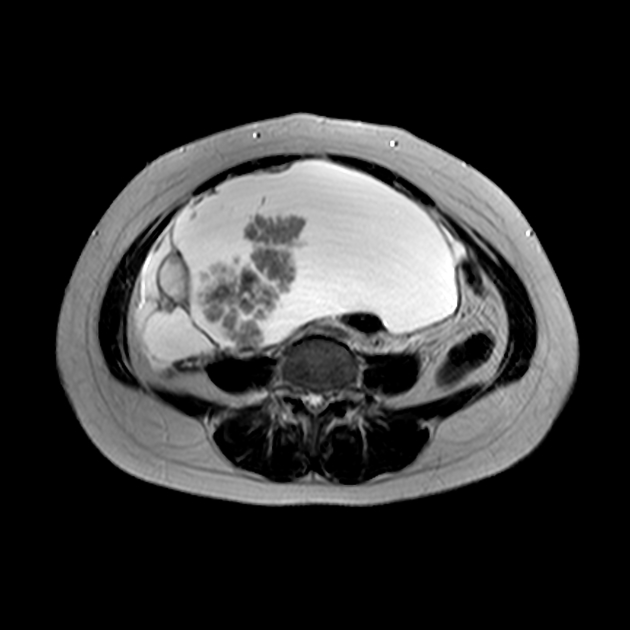
Borderline tumors fall under ovarian epithelial tumors. They tend to develop in an exophytic growth pattern, on the surface of the ovary, without invading the underlying stroma. Papillary projections are characteristic and may be more of a feature with borderline than malignant serous cystadenocarcinoma of the ovary.
A unique feature of borderline tumors is the non-invasive behavior of extra-ovarian tumor implants in the advanced stages of the disease 2-3. Implants can occur in the contralateral ovary, omentum, and peritoneal surface in the advanced stages, although they behave in a benign fashion and remain located on the surface of the underlying tissues.
Markers
Serum CA-125 level is typically mildly elevated.
Radiographic features
Typically seen as bilateral adnexal masses with profuse papillary projections. Bilaterality occurs more frequently than with benign ovarian serous cystadenomas 1.
Serous borderline tumors may display aggressive behavior, and occasionally present with peritoneal or nodal metastases.
Ultrasound
The rate of detection of intratumoral blood flow on Doppler ultrasound can be very similar to more malignant neoplasms 2.
Treatment and prognosis
Post-surgical prognosis is better than for ovarian cystadenocarcinoma, even in the presence of transovarian spread 5.
Staging
Borderline tumors are staged using the same ovarian cancer staging as malignant ovarian neoplasms.
History and etymology
They were first described in 1929 and were designated for separate classification in the early 1970s by the World Health Organization 2.
References
- 1. Nucci MR, Oliva E. Gynecologic Pathology. Churchill Livingstone. (2009) ISBN:0443069204. Read it at Google Books - Find it at Amazon
- 2. Burkholz KJ, Wood BP, Zuppan C. Best cases from the AFIP: Borderline papillary serous tumor of the right ovary. Radiographics. 25 (6): 1689-92. doi:10.1148/rg.256055015 - Pubmed citation
- 3. Seidman JD, Kurman RJ. Ovarian serous borderline tumors: a critical review of the literature with emphasis on prognostic indicators. Hum. Pathol. 2000;31 (5): 539-57. - Pubmed citation
- 4. Prat J, De nictolis M. Serous borderline tumors of the ovary: a long-term follow-up study of 137 cases, including 18 with a micropapillary pattern and 20 with microinvasion. Am. J. Surg. Pathol. 2002;26 (9): 1111-28. Am. J. Surg. Pathol. (link) - Pubmed citation
- 5. Wasnik AP, Menias CO, Platt JF et-al. Multimodality imaging of ovarian cystic lesions: Review with an imaging based algorithmic approach. World J Radiol. 2013;5 (3): 113-25. doi:10.4329/wjr.v5.i3.113 - Free text at pubmed - Pubmed citation
Incoming Links
Related articles: Pathology: Genitourinary
- obstetrics
-
first trimester
- ultrasound findings in early pregnancy
- embryo/fetus
- beta-hCG levels
- confirming intrauterine gestation
- pregnancy of unknown location (PUL)
- first trimester vaginal bleeding
- early structural scan
- aneuploidy testing
-
second trimester
- fetal biometry
- amniotic fluid volume
- fetal morphology assessment
- soft markers
- amnioreduction
- Doppler ultrasound
- nuchal translucency
- 11-13 weeks antenatal scan
- chorionic villus sampling (CVS) and amniocentesis
- other
- placenta
- placental anatomy
- placental developmental abnormalities
- placenta previa
- spectrum of abnormal placental villous adherence
- abnormalities of cord insertion
- abruptio placentae
- placental pathology
- vascular pathologies of placenta
- placental infections
- placental masses
- molar pregnancy
- twin placenta
- miscellaneous
-
first trimester
- gynecology
- acute pelvic pain
- chronic pelvic pain
- uterus
- ovaries
- ovarian follicle
- ovarian torsion
- pelvic inflammatory disease
- ovarian cysts and masses
- paraovarian cyst
- polycystic ovaries
- ovarian hyperstimulation syndrome
- post-hysterectomy ovary
- cervix
- fallopian tube
- other
- male genital tract
- prostate gland
- transrectal ultrasound
- prostate tumors
- infections of the prostate
-
prostatitis
- acute bacterial prostatitis
-
chronic prostatitis
- chronic bacterial prostatitis
- chronic prostatitis and chronic pelvic pain syndrome (CPPS)
- asymptomatic inflammatory prostatitis
- granulomatous prostatitis
- emphysematous prostatitis
- prostatic abscess
-
prostatitis
- benign prostatic hypertrophy
- cystic lesions of the prostate
- prostatic calcification
- prostatic infarction
- testes
-
unilateral testicular lesion
- testicular torsion
- orchitis
- testicular trauma
-
germ cell tumors of the testis
- testicular seminoma
-
non seminomatous germ cell tumors
- mixed germ cell tumor
- yolk sac tumor (endodermal sinus tumor)
- embryonal cell carcinoma
- choriocarcinoma
- testicular teratoma
- testicular epidermoid (teratoma with ectodermal elements only)
- burned out testis tumor
- sex cord / stromal tumors of the testis
- testicular cyst
- testicular lymphoma
- bilateral testicular lesion
- paratesticular lesions
- epididymis
- other
- polyorchidism
- cryptorchidism
- tubular ectasia of the rete testis
- cystadenoma of the rete testis
- testicular sarcoidosis
- testicular tuberculosis
- spermatic cord
- fibrous pseudotumor of the scrotum
- scrotal leiomyosarcoma
- testicular adrenal rest tumors (TARTs)
- tunica vaginalis testis mesothelioma
- splenogonadal fusion
- testicular vasculitis
- abnormal testicular Doppler flow (differential)
-
unilateral testicular lesion
- penis
- prostate gland
- KUB
- kidneys
- normal renal anatomy
- hydronephrosis
- urolithiasis
- renal masses
- renal cystic disease
- renal infection
- vascular
- trauma
- ureter
- normal ureter anatomy
- ureteral stricture
- ureteral dilatation
- ureteral anomalies
- ureteral tumors
- ureteral trauma
- other
- bladder
- kidneys


 Unable to process the form. Check for errors and try again.