
In-phase and out-of-phase sequences
Citation, DOI, disclosures and article data
Citation:
Di Muzio B, Saber M, Bickle I, et al. In-phase and out-of-phase sequences. Reference article, Radiopaedia.org (Accessed on 30 Mar 2025) https://doi.org/10.53347/rID-42534
rID:
42534
Article created:
Disclosures:
At the time the article was created Bruno Di Muzio had no recorded disclosures.
View Bruno Di Muzio's current disclosures
Last revised:
Disclosures:
At the time the article was last revised Mohamed Saber had no recorded disclosures.
View Mohamed Saber's current disclosures
Revisions:
18 times, by
13 contributors -
see full revision history and disclosures
Sections:
Tags:
Synonyms:
- In-phase and out-of-phase sequences
- In-phase/out-of-phase
- IP and OOP
- IP/OOP
- IP-OOP
- In-phase and out-of-phase
- Opposed-phase
- Chemical shift cancellation artifact
- Opposed-phase imaging
- in and out of phase
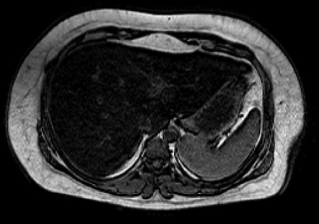
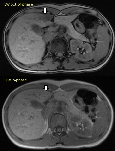
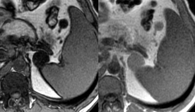
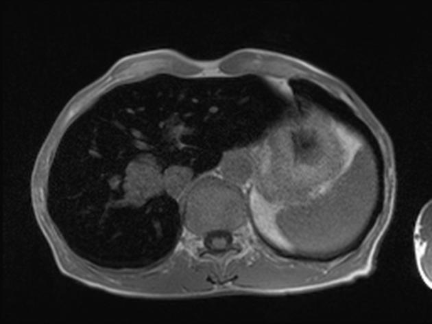
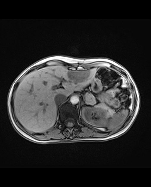
In-phase (IP) and out-of-phase (OOP) sequences correspond to paired MRI gradient echo (GRE) sequences obtained with the same repetition time (TR) but with two different echo time (TE) values.
Applications
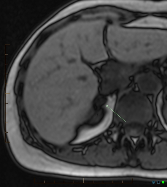
The main application of the IP-OOP sequences is to identify pathological (microscopic) fat content of tissues in the abdomen by showing signal intensities drop on the OOP images compared to the IP images. Loss of signal intensity between the in-phase and out-of-phase MR images indicates fat. Examples where IP-OOP sequences are useful include 1,2:
- fatty liver and focal fatty sparing/infiltration
- fat-rich adrenal lesions:
- adrenal adenoma (helping differentiate it from carcinomas and metastases)
- adrenal myelolipoma
- lipid-poor angiomyolipoma
- renal cell carcinoma (RCC)
- thymic hyperplasia
- osteoporotic versus neoplastic vertebral wedging: neoplastic lesions display persistence high signals on opposed-phase 3,4
- vertebral metastases: metastatic lesions don't show loss of signal in out-of-phase sequence
- hemochromatosis of the liver 5
- intrahepatic pneumobilia 5
- metallic objects: by the effect of susceptibility artifact (e.g cholecystectomy clips)
References
- 1. Ramalho M, Herédia V, de Campos RO et-al. In-phase and out-of-phase gradient-echo imaging in abdominal studies: intra-individual comparison of three different techniques. Acta Radiol. 2012;53 (4): 441-9. doi:10.1258/ar.2012.110695 - Pubmed citation
- 2. Shetty AS, Sipe AL, Zulfiqar M et-al In-Phase and Opposed-Phase Imaging: Applications of Chemical Shift and Magnetic Susceptibility in the Chest and Abdomen. (2019) Radiographics : a review publication of the Radiological Society of North America, Inc. 39 (1): 115-135. doi:10.1148/rg.2019180043 - Pubmed
- 3. Mary Y. Tadros, Amir L. Louka. Discrimination between benign and malignant in vertebral marrow lesions with diffusion weighted MRI and chemical shift. The Egyptian Journal of Radiology and Nuclear Medicine, 47(2), 557-569. https://doi.org/10.1016/j.ejrnm.2016.02.007 - Sciencedirect
- 4. Erly, W., Oh, E., & Outwater, E. The utility of in-phase/opposed-phase imaging in differentiating malignancy from acute benign compression fractures of the spine. (2006) American Journal of Neuroradiology, 27(6), 1183-1188. AJNR
- 5. Merkle EM, Nelson RC. Dual gradient-echo in-phase and opposed-phase hepatic MR imaging: a useful tool for evaluating more than fatty infiltration or fatty sparing. (2006) Radiographics : a review publication of the Radiological Society of North America, Inc. 26 (5): 1409-18. doi:10.1148/rg.265055711 - Pubmed
Incoming Links
Articles:
- Epithelioid angiomyolipoma of kidney
- Thoracic spine protocol (MRI)
- Adrenal metastasis
- Adrenal glands protocol (MRI)
- Pancreatic ductal adenocarcinoma
- Lumbar spine protocol (MRI)
- Perinephric myxoid pseudotumour of fat
- Focal fatty sparing of the liver
- Thymus protocol (MRI)
- Liver protocol (MRI)
- Dixon method
- Adrenal adenoma
- MRI pulse sequence abbreviations
- Medical abbreviations and acronyms (I)
- Aggressive vertebral haemangioma
- Thymus
- Metabolic dysfunction associated steatotic liver disease
- Haemochromatosis (pancreatic manifestations)
- Cervical spine protocol (MRI)
- Medical abbreviations and acronyms (O)
Cases:
- Adrenal adenoma
- Red sausage
- Disc extrusion - interval growth
- Hepatic adenoma
- Adrenal adenoma
- Lipid-rich adrenal adenoma
- Solid pseudopapillary neoplasm of the pancreas
- Fatty liver
- Adrenal adenoma (chemical shift imaging)
- Renal angiomyolipoma
- Angiomyolipoma
- Acute pancreatitis with incidental pancreatic lipoma
- Renal angiomyolipoma
- Haemochromatosis
- Focal fatty change
- Adrenal adenoma
Related articles: Imaging technology
- imaging technology
- imaging physics
- imaging in practice
-
x-rays
- x-ray physics
- x-ray in practice
- x-ray production
- x-ray tube
- filters
- automatic exposure control (AEC)
- beam collimators
- grids
- air gap technique
- cassette
- intensifying screen
- x-ray film
- image intensifier
- digital radiography
- digital image
- mammography
- x-ray artifacts
- radiation units
- radiation safety
- radiation detectors
- fluoroscopy
-
computed tomography (CT)
- CT physics
- CT in practice
- CT technology
- CT image reconstruction
- CT image quality
- CT dose
-
CT contrast media
-
iodinated contrast media
- agents
- water soluble
- water insoluble
- vicarious contrast material excretion
- iodinated contrast media adverse reactions
- agents
- non-iodinated contrast media
-
iodinated contrast media
-
CT artifacts
- patient-based artifacts
- physics-based artifacts
- hardware-based artifacts
- ring artifact
- tube arcing
- out of field artifact
- air bubble artifact
- helical and multichannel artifacts
- CT safety
- history of CT
-
MRI
- MRI physics
- MRI in practice
- MRI hardware
- signal processing
-
MRI pulse sequences (basics | abbreviations | parameters)
- T1 weighted image
- T2 weighted image
- proton density weighted image
- chemical exchange saturation transfer
- CSF flow studies
- diffusion weighted imaging (DWI)
- echo-planar pulse sequences
- fat-suppressed imaging sequences
- gradient echo sequences
- inversion recovery sequences
- metal artifact reduction sequence (MARS)
-
perfusion-weighted imaging
- techniques
- derived values
- saturation recovery sequences
- spin echo sequences
- spiral pulse sequences
- susceptibility-weighted imaging (SWI)
- T1 rho
- MR angiography (and venography)
-
MR spectroscopy (MRS)
- 2-hydroxyglutarate peak: resonates at 2.25 ppm
- alanine peak: resonates at 1.48 ppm
- choline peak: resonates at 3.2 ppm
- citrate peak: resonates at 2.6 ppm
- creatine peak: resonates at 3.0 ppm
- functional MRI (fMRI)
- gamma-aminobutyric acid (GABA) peak: resonates at 2.2-2.4 ppm
- glutamine-glutamate peak: resonates at 2.2-2.4 ppm
- Hunter's angle
- lactate peak: resonates at 1.3 ppm
- lipids peak: resonates at 1.3 ppm
- myoinositol peak: resonates at 3.5 ppm
- MR fingerprinting
- N-acetylaspartate (NAA) peak: resonates at 2.0 ppm
- propylene glycol peak: resonates at 1.13 ppm
-
MRI artifacts
- MRI hardware and room shielding
- MRI software
- patient and physiologic motion
- tissue heterogeneity and foreign bodies
- Fourier transform and Nyquist sampling theorem
- MRI contrast agents
- MRI safety
-
ultrasound
- ultrasound physics
-
transducers
- linear array
- convex array
- phased array
- frame averaging (frame persistence)
- ultrasound image resolution
- imaging modes and display
- pulse-echo imaging
- real-time imaging
-
Doppler imaging
- Doppler effect
- color Doppler
- power Doppler
- B flow
- color box
- Doppler angle
- pulse repetition frequency and scale
- wall filter
- color write priority
- packet size (dwell time)
- peak systolic velocity
- end-diastolic velocity
- resistive index
- pulsatility index
- Reynolds number
- panoramic imaging
- compound imaging
- harmonic imaging
- elastography
- scanning modes
- 2D ultrasound
- 3D ultrasound
- 4D ultrasound
- M-mode
-
ultrasound artifacts
- acoustic shadowing
- acoustic enhancement
- beam width artifact
- reverberation artifact
- ring down artifact
- mirror image artifact
- side lobe artifact
- speckle artifact
- speed displacement artifact
- refraction artifact
- multipath artifact
- anisotropy
- electrical interference artifact
- hardware-related artifacts
- Doppler artifacts
- aliasing
- tissue vibration
- spectral broadening
- blooming
- motion (flash) artifact
- twinkling artifact
- acoustic streaming
- biological effects of ultrasound
- history of ultrasound
-
nuclear medicine
- nuclear medicine physics
- detectors
- tissue to background ratio
-
radiopharmaceuticals
- fundamentals of radiopharmaceuticals
- radiopharmaceutical labeling
- radiopharmaceutical production
- nuclear reactor produced radionuclides
- cyclotron produced radionuclides
- radiation detection
- dosimetry
- specific agents
- carbon-11
- chromium-51
- fluorine agents
- gallium agents
- Ga-67 citrate
- Ga-68
- iodine agents
-
I-123
- I-123 iodide
- I-123 ioflupane (DaTSCAN)
- I-123 ortho-iodohippurate
- I-131
-
MIBG scans
- I-123 MIBG
- I-131 MIBG
-
I-123
- indium agents
- In-111 Octreoscan
- In-111 OncoScint
- In-111 Prostascint
- In-111 oxine labeled WBC
- krypton-81m
- nitrogen-13
- oxygen-15
- phosphorus-32
- selenium-75
-
technetium agents
- Tc-99m DMSA
- Tc-99m DTPA
- Tc-99m DTPA aerosol
- Tc-99m HMPAO
- Tc-99m HMPAO labeled WBC
- Tc-99m MAA
- Tc-99m MAG3
- Tc-99m MDP
- Tc-99m mercaptoacetyltriglycine
- Tc-99m pertechnetate
- Tc-99m labeled RBC
- Tc-99m sestamibi
- Tc-99m sulfur colloid
- Tc-99m sulfur colloid (oral)
- thallium-201 chloride
- xenon agents
- in vivo therapeutic agents
- pharmaceuticals used in nuclear medicine
-
emerging methods in medical imaging
- radiography
- phase-contrast imaging
- CT
- deep-learning reconstruction
- photon counting CT
- virtual non-contrast imaging
- ultrasound
- magnetomotive ultrasound (MMUS)
- superb microvascular imaging
- ultrafast Doppler imaging
- ultrasound localization microscopy
- MRI
- nuclear medicine
- total body PET system
- immuno-PET
- miscellaneous
- radiography


 Unable to process the form. Check for errors and try again.