
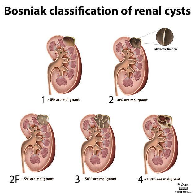
The Bosniak classification system of renal cystic masses, now known as Bosniak v2005, divides renal cystic masses into five categories based on imaging characteristics on contrast-enhanced CT, and helps predict a risk of malignancy and suggests either follow-up or treatment. An updated classification, Bosniak v2019, has been published, and is gradually replacing v2005 in mainstream usage.
On this page:
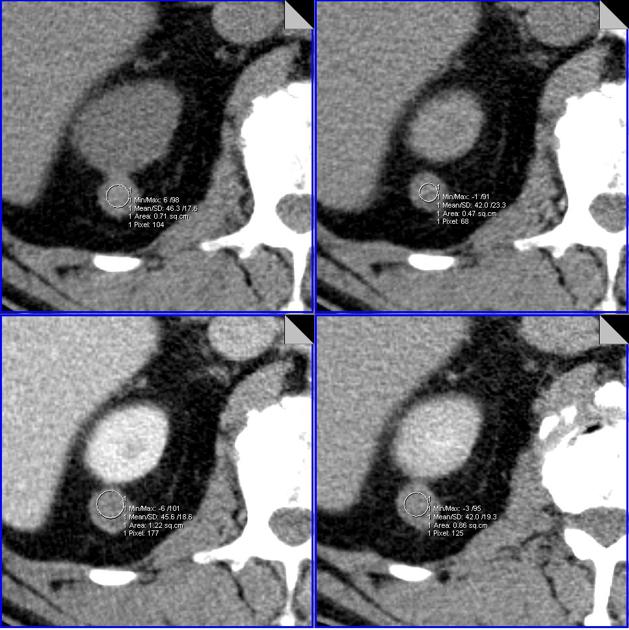
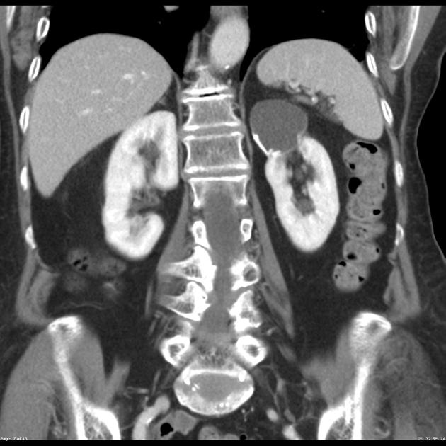
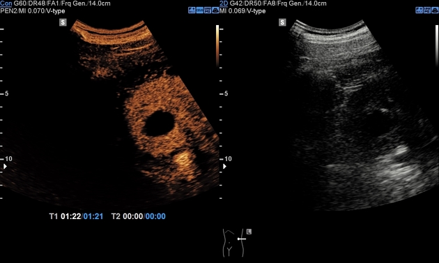
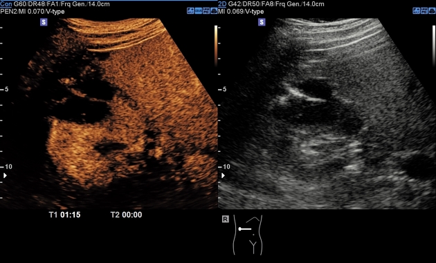
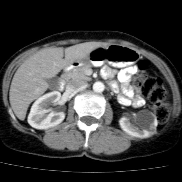
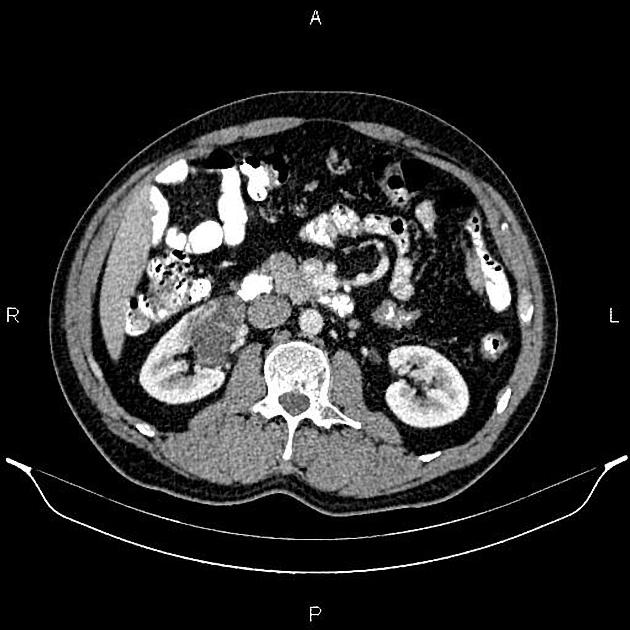
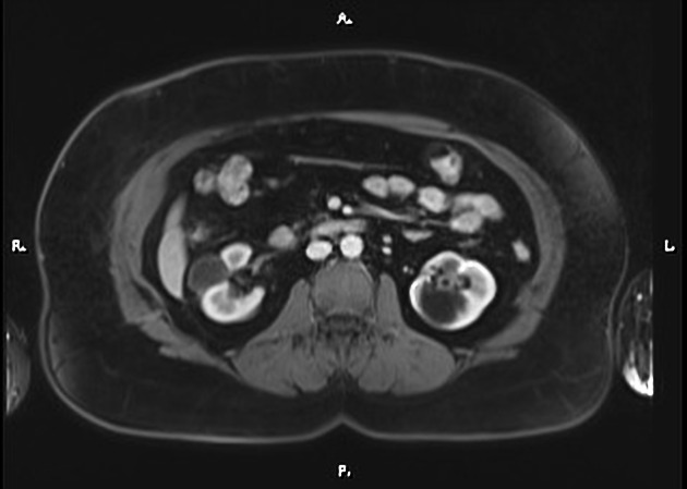
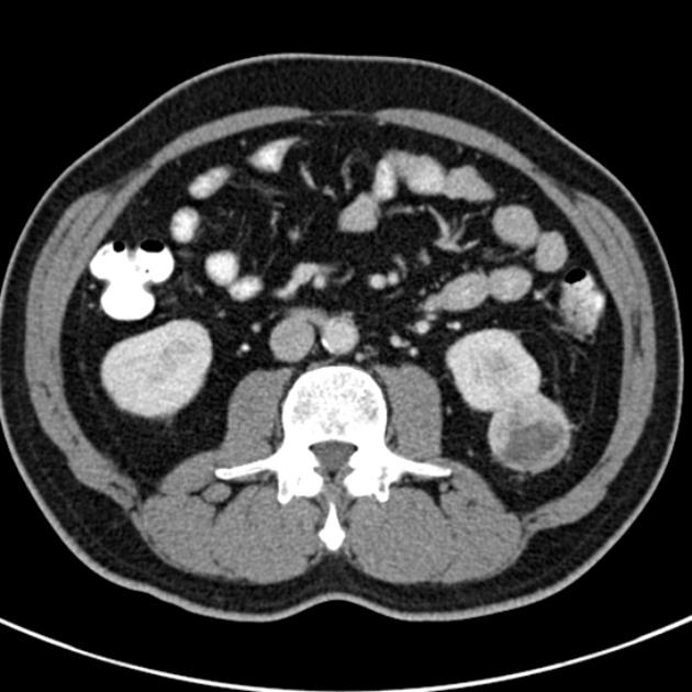
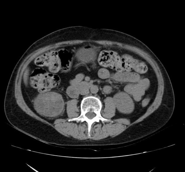
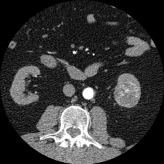
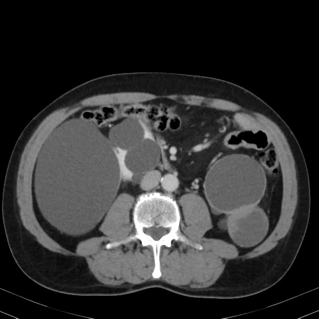
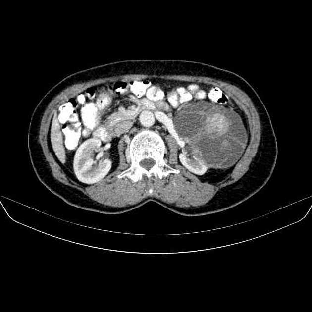
Images:
Usage
Radiologists and urologists widely use the Bosniak classification for assessing renal cysts 3 and was last updated in 2005 12. An updated classification, Bosniak v2019, has been published, including separate CT and MRI-based classification 11.
The Bosniak classification version referred to in international guidelines is variable, with the 2022 European and Canadian guidelines referencing v2019 25,26 and the 2021 American guidelines referencing v2005 27,28.
Although practised by some, using ultrasonography to characterize the Bosniak classification remains controversial. Originally, it was thought ultrasound was inadequate for the task as it could not show neovascularization (cf. contrast-enhanced CT/MRI) ref. However, newer studies looking at contrast-enhanced ultrasound suggest this impediment is no longer true ref. There is also evidence that ultrasound has a higher sensitivity for intralesional septa than either CT or MRI 8,13.
Classification
The "official" Bosniak classification uses Roman numerals, not Arabic ones, for each category. The use of the term "grade", "stage", "group", "type", or similar for each category is technically incorrect. Version 2019 has switched from "category" to "class" 11.
Bosniak I
-
benign simple cyst
hairline-thin wall of ≤2 mm
water density
no septa, calcifications, or solid components
no enhancement
work-up: none
percentage malignant: ~0% 17
Bosniak II
-
benign cyst - "minimally complex"
few hairlines thin <1 mm septa or thin calcifications (thickness not measurable)
perceived enhancement
non-enhancing high-attenuation (due to proteinaceous or hemorrhagic contents) renal lesions <3 cm
generally well marginated
work-up: none
percentage malignant: ~0-6% 17,18
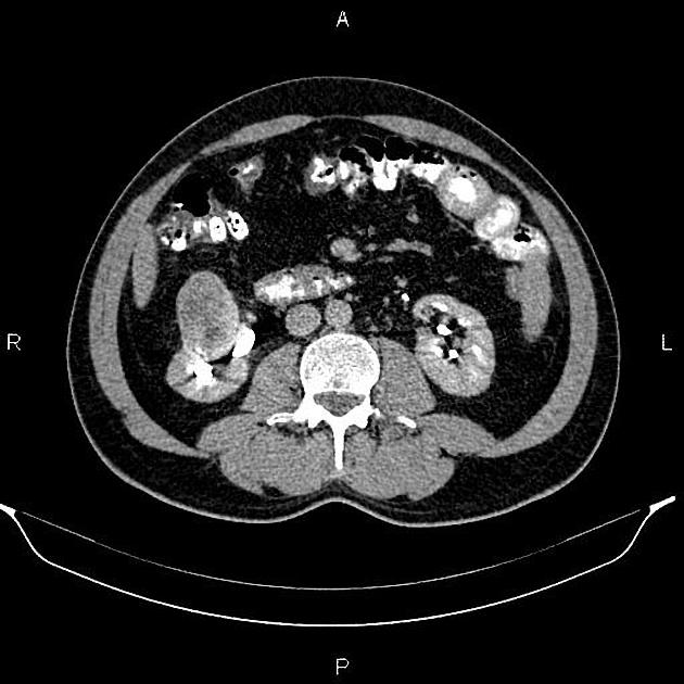
Bosniak IIF
-
minimally complex
multiple hairline thin septa or minimally smooth thickened walls or septa
perceived but no measurable enhancement of wall or septa
calcification can be present and may be thick and nodular
generally well marginated
high-attenuation lesion >3 cm diameter, totally intrarenal (<25% of wall visible); no enhancement
requiring follow-up (F for follow-up): needs ultrasound/CT/MRI follow up - no strict rules on the time frame but reasonable at 6 months, 12 months, then annually for 5 years 3
percentage malignant: ~5-26% 6,19-21
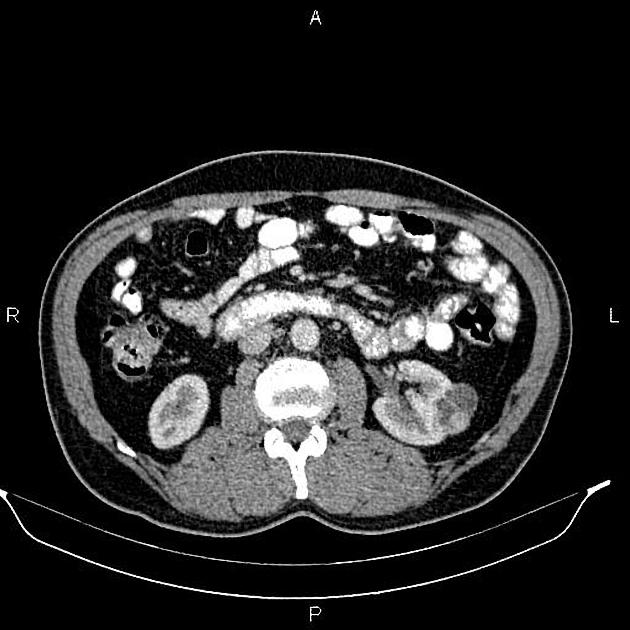
Bosniak III
-
indeterminate cystic mass
thickened irregular or smooth walls or septa with measurable enhancement
treatment/work-up: partial nephrectomy or radiofrequency ablation in poor surgical candidates 23,24
percentage malignant: ~55-72% 6,17,19,22
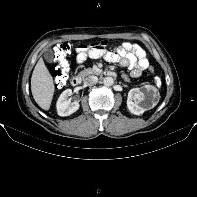
Bosniak IV
-
clearly malignant cystic mass
Bosniak III criteria + enhancing soft tissue components adjacent to but independent of wall or septum
treatment: partial or total nephrectomy
percentage malignant: ~91-100% 19,22
History and etymology
The Bosniak classification is named after Morton A Bosniak (1929-2016), who was professor emeritus in radiology at New York University (NYU) Langone School of Medicine. It was first published in 1986, introducing the 2F category in 1993, and revisions in 1997, 2005 and 2019 9,10,14-16.


 Unable to process the form. Check for errors and try again.