Discoid menisci are anatomical variants that have a body that is too wide, usually affecting the lateral meniscus. They are incidentally found in 3-5% of knee MRI examinations.
On this page:
Epidemiology
Discoid menisci are congenital, frequently bilateral (up to 50%) and have been reported in twins, although no genetic locus has been identified 2. There is a higher prevalence in Asians without any gender predilection 7. Lateral discoid meniscus is far more common than medial discoid meniscus, with the latter being rare.
Clinical presentation
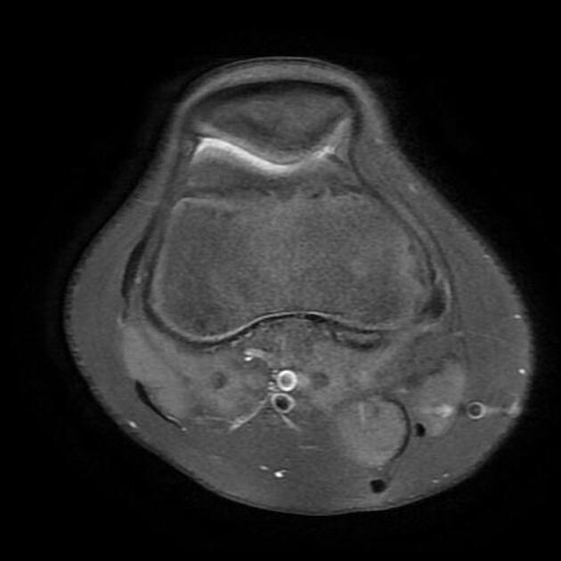
Although frequently asymptomatic, discoid menisci are prone to cystic degeneration with a subsequent tear. Clinical presentation may, therefore, be either incidental or with pain, locking or a 'clunk'.
Pathology
Discoid menisci have decreased collagen fibers and loss of normal collagen orientation, which predisposes them to intradiscal/meniscal mucoid degeneration 7.
Classification
Classification is based on the degree of peripheral attachments to the tibial plateau, and the shape of the meniscus itself:
-
complete vs incomplete
80% coverage of the tibial plateau is often used as the cut-off between incomplete and complete 7
-
stable vs unstable
stable: normal peripheral attachments with an intact posterior meniscofemoral ligament
unstable (also known as a Wrisberg variant): lack or tear of a posterior meniscocapsular (in particular meniscopopliteal) ligaments with an attachment only from the meniscofemoral ligament of Wrisberg 4
anterior or posterior mega horn
Radiographic features
Plain radiograph
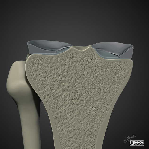
Radiographs may well suggest the diagnosis with the widening of the lateral joint space and cupping of the lateral tibial plateau, which is normally flat or even slightly convex. Additionally, there may be associated hypoplasia of the lateral tibial spine.
MRI
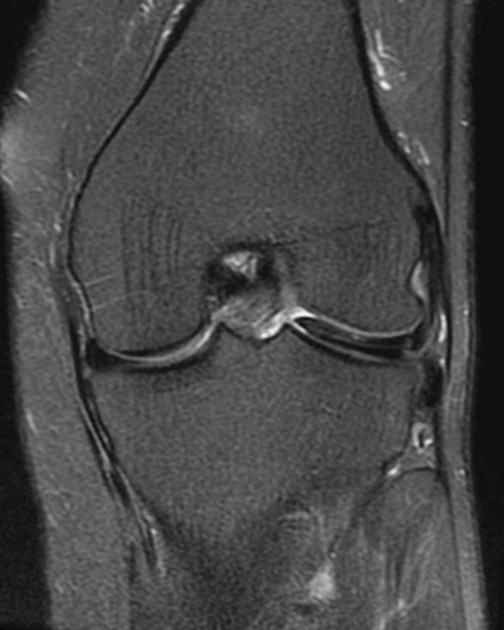
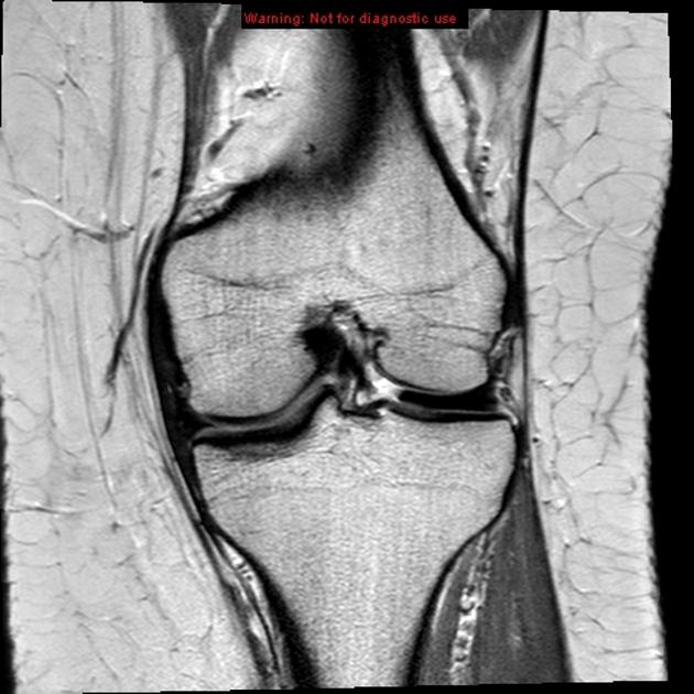
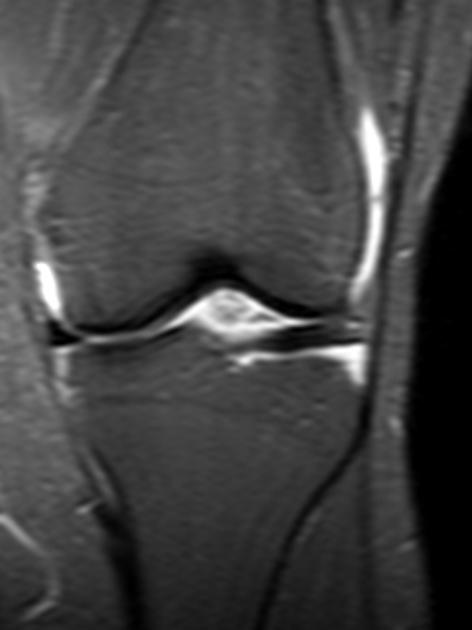
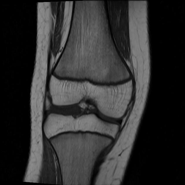
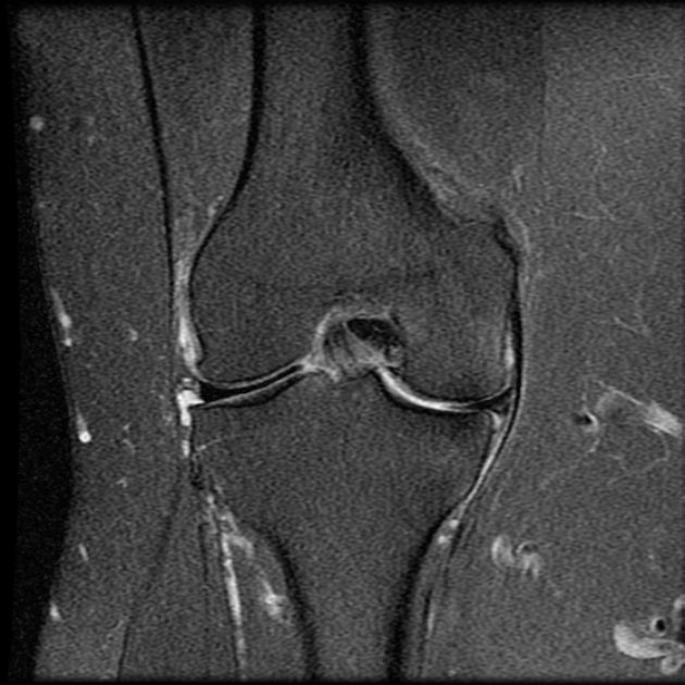
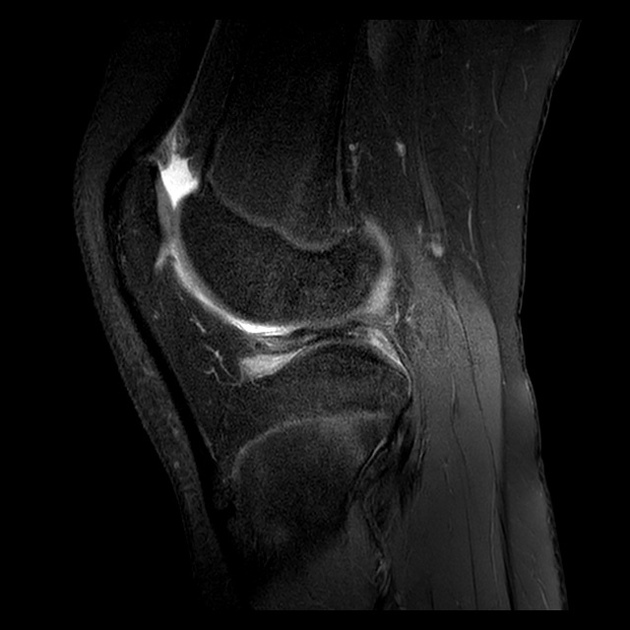
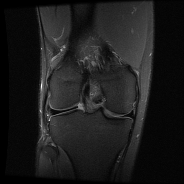
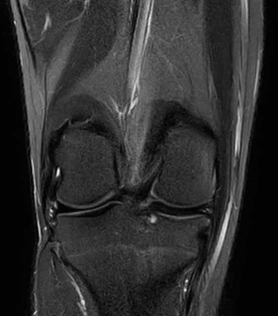
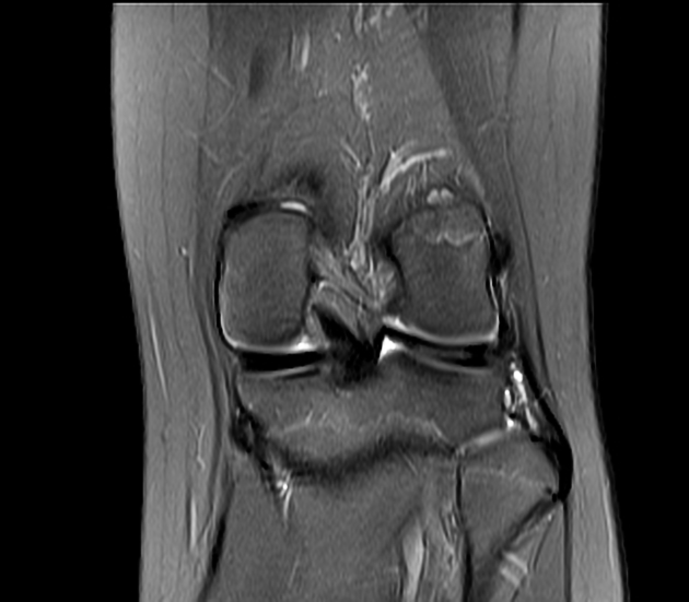
On coronal imaging, meniscal body width of 15 mm or more is typically considered diagnostic of discoid morphology. Alternatively, a ratio of the minimal meniscal width to the maximal tibial width of more than 20% may be used 7,8.
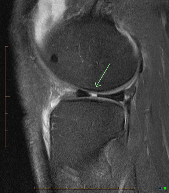
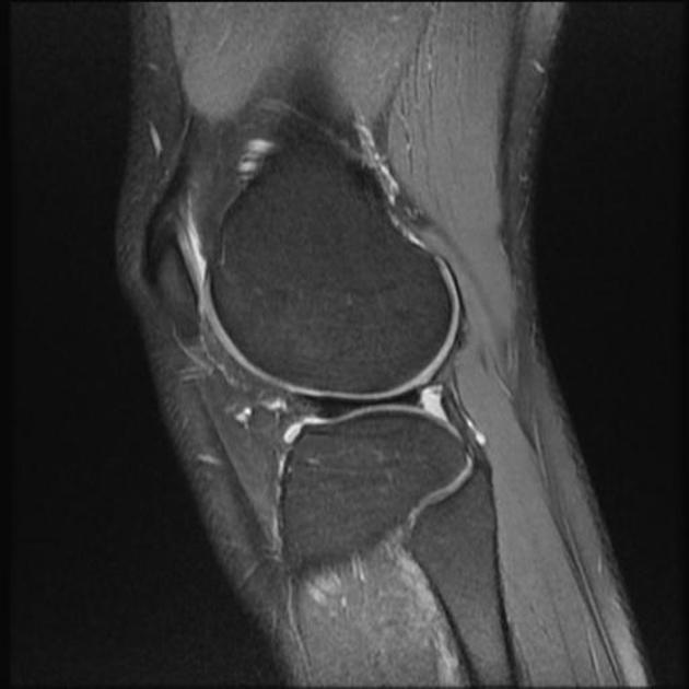
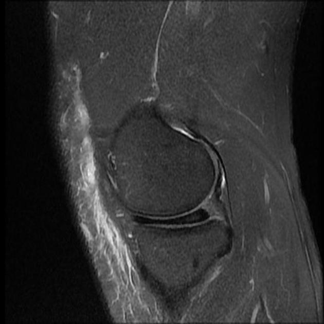
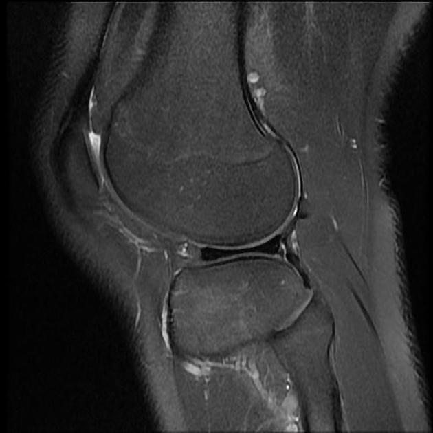
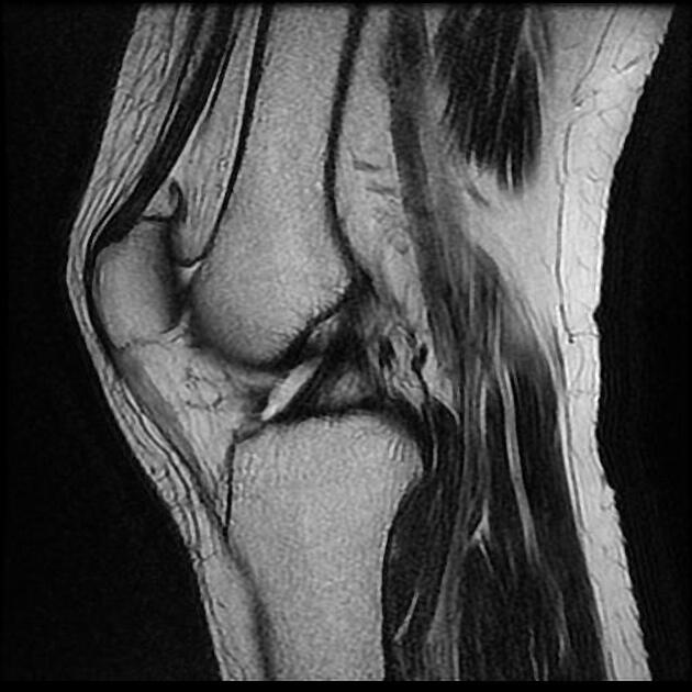
On sagittal imaging, the body of the lateral meniscus is normally only seen on two adjacent slices. A discoid meniscus is usually present if the meniscal body is seen on three or more standard sagittal slices - the opposite of the absent bow tie sign.
Treatment and prognosis
Ideally, the meniscal rim is preserved with conservative management. If this is unsuccessful, then meniscal repair with partial or total resection may be carried out 09.
Complications
intrasubstance mucoid degeneration
early bony degenerative change






 Unable to process the form. Check for errors and try again.