
Cervical ectopic pregnancy
Citation, DOI, disclosures and article data
At the time the article was created Frank Gaillard had no recorded disclosures.
View Frank Gaillard's current disclosuresAt the time the article was last revised Jeremy Jones had no recorded disclosures.
View Jeremy Jones's current disclosures- Cervical ectopic
- Cervical ectopic pregnancies
- Cervical ectopics
Cervical ectopic pregnancy is a rare subtype of ectopic pregnancy, in which a gestational sac is seen at the endocervical canal below the closed internal os.
On this page:
Epidemiology
It accounts for ~0.15-1% of all ectopic pregnancies.
Pathology
Implantation of the fertilized ovum occurs within the cervix rather than the uterine cavity. Unless the fetal heart rate can be identified, it is difficult to distinguish from a miscarriage with fetal parts in the cervical os.
Risk factors
- variant anatomy 10
- fibroids 10
- IUCD
- Asherman syndrome
- IVF
- repeated dilatation and curettage 8
Radiographic assessment
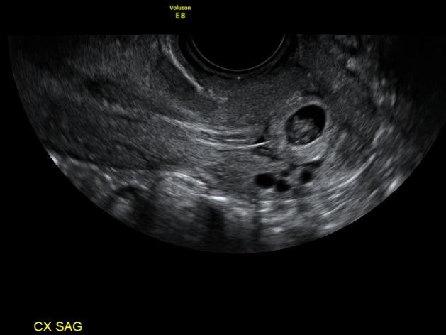
Ultrasound
Can be seen as a gestational sac within the distended cervix which gives an hour-glass appearance to the uterus. Usually, internal os is closed. At times the gestational sac extends into the lower uterine segment (abnormally low sac position). There is hyperechoic decidual reaction around the gestational sac.
Color Doppler imaging can be helpful in showing a hypervascular trophoblastic ring in the cervical region in cases of live cervical ectopic pregnancies.
Treatment and prognosis
The goal is to treat the condition whilst minimizing the risk of severe hemorrhage, and to preserve the patient's future reproductive potential.
Medical management options include methotrexate (a folate antagonist) either administered systemically or by direct injection, or potassium chloride (direct injection).
Surgical curettage runs the risk of life-threatening hemorrhage. Preoperative uterine embolization is an option for reduction of hemorrhage risk 4,6,7.
Complications
Severe hemorrhage is the main complication of surgical management. It may result in the need for hysterectomy, and is potentially life-threatening.
Differential diagnosis
- it is important to consider miscarriage in progress as a differential when the gestational sac is located within the endocervical canal; features suggestive of a miscarriage include:
- absent embryonic cardiac activity
- open internal os: lack of typical hourglass appearance
- sac shape and location often changes on serial scans and it may be possible to alter the location of the sac with gentle probe maneuvering ("sliding sac sign")
- subsequent loss of the fetal sac on a repeat ultrasound confirms miscarriage
References
- 1. Weissleder R, Wittenberg J, Harisinghani MM et-al. Primer of Diagnostic Imaging, Expert Consult- Online and Print. Mosby. (2011) ISBN:0323065384. Read it at Google Books - Find it at Amazon
- 2. Chudleigh P, Thilaganathan B, Chudleigh T. Obstetric ultrasound, how, why and when. Churchill Livingstone. (2004) ISBN:0443054711. Read it at Google Books - Find it at Amazon
- 3. Merz E, Bahlmann F. Ultrasound in obstetrics and gynecology. Thieme Medical Publishers. (2005) ISBN:1588901475. Read it at Google Books - Find it at Amazon
- 4. Levine D. Ectopic pregnancy. Radiology. 2007;245 (2): 385-97. doi:10.1148/radiol.2452061031 - Pubmed citation
- 5. Leeman LM, Wendland CL. Cervical ectopic pregnancy. Diagnosis with endovaginal ultrasound examination and successful treatment with methotrexate. Arch Fam Med. 2000;9 (1): 72-7. doi:10.1001/archfami.9.1.72 - Pubmed citation
- 6. Frates MC, Benson CB, Doubilet PM et-al. Cervical ectopic pregnancy: results of conservative treatment. Radiology. 1994;191 (3): 773-5. Radiology (abstract) - Pubmed citation
- 7. Meyerovitz MF, Lobel SM, Harrington DP et-al. Preoperative uterine artery embolization in cervical pregnancy. J Vasc Interv Radiol. 1991;2 (1): 95-7. - Pubmed citation
- 8. Chukus A, Tirada N, Restrepo R et-al. Uncommon Implantation Sites of Ectopic Pregnancy: Thinking beyond the Complex Adnexal Mass. Radiographics. 2015;35 (3): 946-59. doi:10.1148/rg.2015140202 - Pubmed citation
- 9. Papaloucas CD. "Hour-glass" shape of the uterus in the diagnosis and treatment of cervical pregnancy. Clin Anat. 2004;17 (8): 658-61. doi:10.1002/ca.10223 - Pubmed citation
- 10. Dibble EH, Lourenco AP. Imaging Unusual Pregnancy Implantations: Rare Ectopic Pregnancies and More. AJR Am J Roentgenol. 2016; 1-13. doi:10.2214/AJR.15.15290 - Pubmed citation
Incoming Links
Related articles: Pathology: Genitourinary
- obstetrics
-
first trimester
- ultrasound findings in early pregnancy
- embryo/fetus
- beta-hCG levels
- confirming intrauterine gestation
- pregnancy of unknown location (PUL)
- first trimester vaginal bleeding
- early structural scan
- aneuploidy testing
-
second trimester
- fetal biometry
- amniotic fluid volume
- fetal morphology assessment
- soft markers
- amnioreduction
- Doppler ultrasound
- nuchal translucency
- 11-13 weeks antenatal scan
- chorionic villus sampling (CVS) and amniocentesis
- other
- placenta
- placental anatomy
- placental developmental abnormalities
- placenta previa
- spectrum of abnormal placental villous adherence
- abnormalities of cord insertion
- abruptio placentae
- placental pathology
- vascular pathologies of placenta
- placental infections
- placental masses
- molar pregnancy
- twin placenta
- miscellaneous
-
first trimester
- gynecology
- acute pelvic pain
- chronic pelvic pain
- uterus
- ovaries
- ovarian follicle
- ovarian torsion
- pelvic inflammatory disease
- ovarian cysts and masses
- paraovarian cyst
- polycystic ovaries
- ovarian hyperstimulation syndrome
- post-hysterectomy ovary
- cervix
- fallopian tube
- other
- male genital tract
- prostate gland
- transrectal ultrasound
- prostate tumors
- infections of the prostate
-
prostatitis
- acute bacterial prostatitis
-
chronic prostatitis
- chronic bacterial prostatitis
- chronic prostatitis and chronic pelvic pain syndrome (CPPS)
- asymptomatic inflammatory prostatitis
- granulomatous prostatitis
- emphysematous prostatitis
- prostatic abscess
-
prostatitis
- benign prostatic hypertrophy
- cystic lesions of the prostate
- prostatic calcification
- prostatic infarction
- testes
-
unilateral testicular lesion
- testicular torsion
- orchitis
- testicular trauma
-
germ cell tumors of the testis
- testicular seminoma
-
non seminomatous germ cell tumors
- mixed germ cell tumor
- yolk sac tumor (endodermal sinus tumor)
- embryonal cell carcinoma
- choriocarcinoma
- testicular teratoma
- testicular epidermoid (teratoma with ectodermal elements only)
- burned out testis tumor
- sex cord / stromal tumors of the testis
- testicular cyst
- testicular lymphoma
- bilateral testicular lesion
- paratesticular lesions
- epididymis
- other
- polyorchidism
- cryptorchidism
- tubular ectasia of the rete testis
- cystadenoma of the rete testis
- testicular sarcoidosis
- testicular tuberculosis
- spermatic cord
- fibrous pseudotumor of the scrotum
- scrotal leiomyosarcoma
- testicular adrenal rest tumors (TARTs)
- tunica vaginalis testis mesothelioma
- splenogonadal fusion
- testicular vasculitis
- abnormal testicular Doppler flow (differential)
-
unilateral testicular lesion
- penis
- prostate gland
- KUB
- kidneys
- normal renal anatomy
- hydronephrosis
- urolithiasis
- renal masses
- renal cystic disease
- renal infection
- vascular
- trauma
- ureter
- normal ureter anatomy
- ureteral stricture
- ureteral dilatation
- ureteral anomalies
- ureteral tumors
- ureteral trauma
- other
- bladder
- kidneys


 Unable to process the form. Check for errors and try again.