
Scrotolith
Citation, DOI, disclosures and article data
At the time the article was created Yuranga Weerakkody had no recorded disclosures.
View Yuranga Weerakkody's current disclosuresAt the time the article was last revised Daniel J Bell had no financial relationships to ineligible companies to disclose.
View Daniel J Bell's current disclosures- Scrotal pearls
- Scrotoliths
- Scrotal calculi
- Scrotal calculus
- Scrotal stone
- Scrotal stones
Scrotoliths, also known as scrotal pearls, are benign incidental extratesticular macrocalcifications within the scrotum. They frequently occupy the potential space of the tunica vaginalis or sinus of the epididymis. They are usually of no clinical significance 1,2.
On this page:
Epidemiology
The prevalence of scrotoliths has been estimated to be approximately 3% 3.
Clinical presentation
Most scrotoliths are asymptomatic.
Pathology
Possible etiologies include:
reactive fibrous proliferation during intrascrotal inflammation microtrauma to the scrotal region, e.g. mountain bikers
calcified loose bodies from prior torsion of the testicular appendix
Radiographic features
Ultrasound
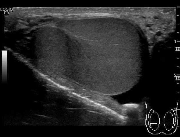
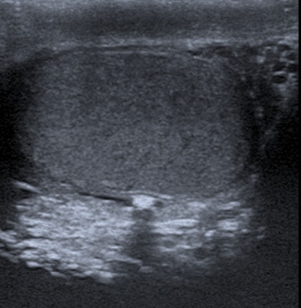
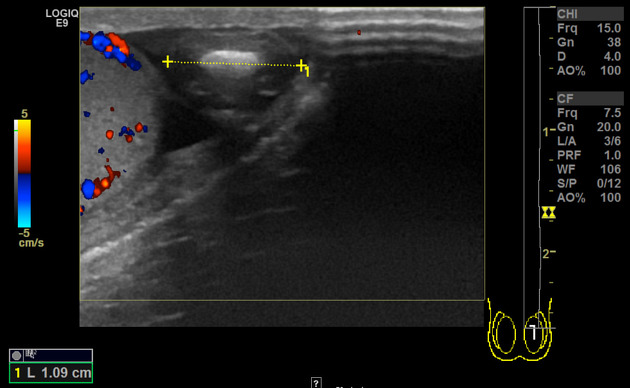
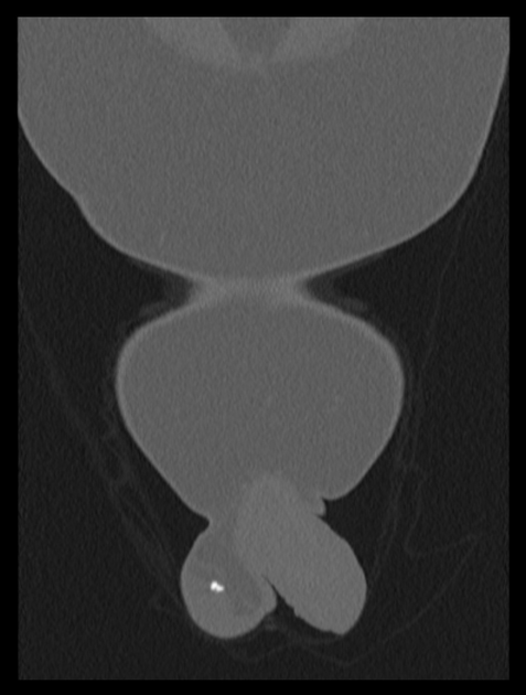
Ultrasound usually shows a small mobile hyperechoic extratesticular focus in the potential tunica space. If large enough, the pearl usually has posterior acoustic shadowing. It may be free floating if there is an accompanying hydrocele.
Most scrotoliths are less than 1 cm in size 6. However occasionally they can be substantially larger, up to 8.7 cm has been reported (in 2024) 7.
References
- 1. Dogra VS, Gottlieb RH, Oka M, Rubens DJ. Sonography of the scrotum. (2003) Radiology. 227 (1): 18-36. doi:10.1148/radiol.2271001744 - Pubmed
- 2. Black JA, Patel A. Sonography of the abnormal extratesticular space. (1996) AJR. American journal of roentgenology. 167 (2): 507-11. doi:10.2214/ajr.167.2.8686637 - Pubmed
- 3. Artas H, Orhan I. Scrotal calculi. (2007) Journal of ultrasound in medicine : official journal of the American Institute of Ultrasound in Medicine. 26 (12): 1775-9. doi:10.7863/jum.2007.26.12.1775 - Pubmed
- 4. Kühn AL, Scortegagna E, Nowitzki KM, Kim YH. Ultrasonography of the scrotum in adults. (2016) Ultrasonography (Seoul, Korea). 35 (3): 180-97. doi:10.14366/usg.15075 - Pubmed
- 5. Mittal PK, Abdalla AS, Chatterjee A, Baumgarten DA, Harri PA, Patel J, Moreno CC, Gabriel H, Miller FH. Spectrum of Extratesticular and Testicular Pathologic Conditions at Scrotal MR Imaging. (2018) Radiographics : a review publication of the Radiological Society of North America, Inc. 38 (3): 806-830. doi:10.1148/rg.2018170150 - Pubmed
- 6. Borthakur D, Dada R, Kumar R, Jacob T. Scrotoliths in the Testicular Tunica Vaginalis in an Elderly Male Cadaver: Clinical Implications. FM. 2024;66(4):574-7. doi:10.3897/folmed.66.e121058 - Pubmed
- 7. Li G, Li X, Cheng S, Chen Z. A Large Intrascrotal Calculus. Asian J Androl. 2005;7(1):103-5. doi:10.1111/j.1745-7262.2005.00013.x - Pubmed
Incoming Links
Related articles: Pathology: Genitourinary
- obstetrics
-
first trimester
- ultrasound findings in early pregnancy
- embryo/fetus
- beta-hCG levels
- confirming intrauterine gestation
- pregnancy of unknown location (PUL)
- first trimester vaginal bleeding
- early structural scan
- aneuploidy testing
-
second trimester
- fetal biometry
- amniotic fluid volume
- fetal morphology assessment
- soft markers
- amnioreduction
- Doppler ultrasound
- nuchal translucency
- 11-13 weeks antenatal scan
- chorionic villus sampling (CVS) and amniocentesis
- other
- placenta
- placental anatomy
- placental developmental abnormalities
- placenta previa
- spectrum of abnormal placental villous adherence
- abnormalities of cord insertion
- abruptio placentae
- placental pathology
- vascular pathologies of placenta
- placental infections
- placental masses
- molar pregnancy
- twin placenta
- miscellaneous
-
first trimester
- gynecology
- acute pelvic pain
- chronic pelvic pain
- uterus
- ovaries
- ovarian follicle
- ovarian torsion
- pelvic inflammatory disease
- ovarian cysts and masses
- paraovarian cyst
- polycystic ovaries
- ovarian hyperstimulation syndrome
- post-hysterectomy ovary
- cervix
- fallopian tube
- other
- male genital tract
- prostate gland
- transrectal ultrasound
- prostate tumors
- infections of the prostate
-
prostatitis
- acute bacterial prostatitis
-
chronic prostatitis
- chronic bacterial prostatitis
- chronic prostatitis and chronic pelvic pain syndrome (CPPS)
- asymptomatic inflammatory prostatitis
- granulomatous prostatitis
- emphysematous prostatitis
- prostatic abscess
-
prostatitis
- benign prostatic hypertrophy
- cystic lesions of the prostate
- prostatic calcification
- prostatic infarction
- testes
-
unilateral testicular lesion
- testicular torsion
- orchitis
- testicular trauma
-
germ cell tumors of the testis
- testicular seminoma
-
non seminomatous germ cell tumors
- mixed germ cell tumor
- yolk sac tumor (endodermal sinus tumor)
- embryonal cell carcinoma
- choriocarcinoma
- testicular teratoma
- testicular epidermoid (teratoma with ectodermal elements only)
- burned out testis tumor
- sex cord / stromal tumors of the testis
- testicular cyst
- testicular lymphoma
- bilateral testicular lesion
- paratesticular lesions
- epididymis
- other
- polyorchidism
- cryptorchidism
- tubular ectasia of the rete testis
- cystadenoma of the rete testis
- testicular sarcoidosis
- testicular tuberculosis
- spermatic cord
- fibrous pseudotumor of the scrotum
- scrotal leiomyosarcoma
- testicular adrenal rest tumors (TARTs)
- tunica vaginalis testis mesothelioma
- splenogonadal fusion
- testicular vasculitis
- abnormal testicular Doppler flow (differential)
-
unilateral testicular lesion
- penis
- prostate gland
- KUB
- kidneys
- normal renal anatomy
- hydronephrosis
- urolithiasis
- renal masses
- renal cystic disease
- renal infection
- vascular
- trauma
- ureter
- normal ureter anatomy
- ureteral stricture
- ureteral dilatation
- ureteral anomalies
- ureteral tumors
- ureteral trauma
- other
- bladder
- kidneys


 Unable to process the form. Check for errors and try again.