
Octreotide scintigraphy
Citation, DOI, disclosures and article data
At the time the article was created Yuranga Weerakkody had no recorded disclosures.
View Yuranga Weerakkody's current disclosuresAt the time the article was last revised Yahya Baba had no financial relationships to ineligible companies to disclose.
View Yahya Baba's current disclosures- Octreotide scan
- In-111 Octreoscan
- Octreoscan
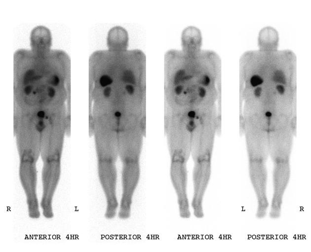
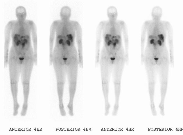
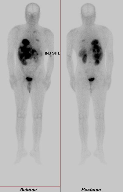
Octreotide scintigraphy uses 111In-labeled octreotide, which is a somatostatin analog; it is also known as Octreoscan, a brand name for 111In-labeled pentetreotide. Pentetreotide is a DTPA-conjugated form of octreotide, originally manufactured by Mallinckrodt Nuclear Medicine LLC, which now forms part of Curium.
The radiotracer is characterized by a high affinity of bond with the somatostatin receptor 2 (SST2). It penetrates inside the cell by endocytosis, first held by lysosomes and then transferred to the nucleus 4,5.
It is particularly useful for the assessment of neuroendocrine tumors, and the Krenning Score is used to grade these, most commonly for peptide receptor radionuclide therapy (PRRT) planning.
Examples include:
Certain non-neuroendocrine tumors can also show uptake in an Octreoscan. Examples include:
Also, certain autoimmune or granulomatous diseases (for example sarcoidosis) may occasionally show octreotide uptake by overexpression of somatostatin receptors 6-8.
Quiz questions
References
- 1. Whiteman M, Serafini A, Telischi F, Civantos F, Falcone S. 111In Octreotide Scintigraphy in the Evaluation of Head and Neck Lesions. AJNR Am J Neuroradiol. 1997;18(6):1073-80. PMC8337293 - Pubmed
- 2. Ramage J, Williams R, Buxton-Thomas M. Imaging Secondary Neuroendocrine Tumours of the Liver: Comparison of I123 Metaiodobenzylguanidine (MIBG) and In111-Labelled Octreotide (Octreoscan). QJM. 1996;89(7):539-42. doi:10.1093/qjmed/89.7.539 - Pubmed
- 3. Guitera-Rovel P, Lumbroso J, Gautier-Gougis M et al. Indium-111 Octreotide Scintigraphy of Merkel Cell Carcinomas and Their Metastases. Ann Oncol. 2001;12(6):807-11. doi:10.1023/a:1011142410535 - Pubmed
- 4. Hornick C, Anthony C, Hughey S, Gebhardt B, Espenan G, Woltering E. Progressive Nuclear Translocation of Somatostatin Analogs. J Nucl Med. 2000;41(7):1256-63. - Pubmed
- 5. Janson E, Westlin J, Ohrvall U, Oberg K, Lukinius A. Nuclear Localization of 111In After Intravenous Injection of [111In-DTPA-D-Phe1]-Octreotide in Patients with Neuroendocrine Tumors. J Nucl Med. 2000;41(9):1514-8. - Pubmed
- 6. Dalm V, van Hagen P, Krenning E. The Role of Octreotide Scintigraphy in Rheumatoid Arthritis and Sarcoidosis. Q J Nucl Med. 2003;47(4):270-8. - Pubmed
- 7. van Hagen P, Krenning E, Kwekkeboom D et al. Somatostatin and the Immune and Haematopoetic System; a Review. Eur J Clin Invest. 1994;24(2):91-9. doi:10.1111/j.1365-2362.1994.tb00972.x - Pubmed
- 8. ten Bokum A, Hofland L, van Hagen P. Somatostatin and Somatostatin Receptors in the Immune System: A Review. Eur Cytokine Netw. 2000;11(2):161-76. - Pubmed
Incoming Links
- Pancreatic neuroendocrine tumours
- Carotid body tumor
- Gastrinoma
- Gastrointestinal neuroendocrine tumours
- Phaeochromocytoma
- Incidental thyroid nodule
- Medical abbreviations and acronyms (S)
- Carcinoid tumour
- Lutetium-177 dotatate
- Paragangliomas of the head and neck
- Gallium-68 DOTATATE
- Bronchial carcinoid tumour
- Krenning score of neuroendocrine tumour uptake
Related articles: Imaging technology
- imaging technology
- imaging physics
- imaging in practice
-
x-rays
- x-ray physics
- x-ray in practice
- x-ray production
- x-ray tube
- filters
- automatic exposure control (AEC)
- beam collimators
- grids
- air gap technique
- cassette
- intensifying screen
- x-ray film
- image intensifier
- digital radiography
- digital image
- mammography
- x-ray artifacts
- radiation units
- radiation safety
- radiation detectors
- fluoroscopy
-
computed tomography (CT)
- CT physics
- CT in practice
- CT technology
- CT image reconstruction
- CT image quality
- CT dose
-
CT contrast media
-
iodinated contrast media
- agents
- water soluble
- water insoluble
- vicarious contrast material excretion
- iodinated contrast media adverse reactions
- agents
- non-iodinated contrast media
-
iodinated contrast media
-
CT artifacts
- patient-based artifacts
- physics-based artifacts
- hardware-based artifacts
- ring artifact
- tube arcing
- out of field artifact
- air bubble artifact
- helical and multichannel artifacts
- CT safety
- history of CT
-
MRI
- MRI physics
- MRI in practice
- MRI hardware
- signal processing
-
MRI pulse sequences (basics | abbreviations | parameters)
- T1 weighted image
- T2 weighted image
- proton density weighted image
- chemical exchange saturation transfer
- CSF flow studies
- diffusion weighted imaging (DWI)
- echo-planar pulse sequences
- fat-suppressed imaging sequences
- gradient echo sequences
- inversion recovery sequences
- metal artifact reduction sequence (MARS)
-
perfusion-weighted imaging
- techniques
- derived values
- saturation recovery sequences
- spin echo sequences
- spiral pulse sequences
- susceptibility-weighted imaging (SWI)
- T1 rho
- MR angiography (and venography)
-
MR spectroscopy (MRS)
- 2-hydroxyglutarate peak: resonates at 2.25 ppm
- alanine peak: resonates at 1.48 ppm
- choline peak: resonates at 3.2 ppm
- citrate peak: resonates at 2.6 ppm
- creatine peak: resonates at 3.0 ppm
- functional MRI (fMRI)
- gamma-aminobutyric acid (GABA) peak: resonates at 2.2-2.4 ppm
- glutamine-glutamate peak: resonates at 2.2-2.4 ppm
- Hunter's angle
- lactate peak: resonates at 1.3 ppm
- lipids peak: resonates at 1.3 ppm
- myoinositol peak: resonates at 3.5 ppm
- MR fingerprinting
- N-acetylaspartate (NAA) peak: resonates at 2.0 ppm
- propylene glycol peak: resonates at 1.13 ppm
-
MRI artifacts
- MRI hardware and room shielding
- MRI software
- patient and physiologic motion
- tissue heterogeneity and foreign bodies
- Fourier transform and Nyquist sampling theorem
- MRI contrast agents
- MRI safety
-
ultrasound
- ultrasound physics
-
transducers
- linear array
- convex array
- phased array
- frame averaging (frame persistence)
- ultrasound image resolution
- imaging modes and display
- pulse-echo imaging
- real-time imaging
-
Doppler imaging
- Doppler effect
- color Doppler
- power Doppler
- B flow
- color box
- Doppler angle
- pulse repetition frequency and scale
- wall filter
- color write priority
- packet size (dwell time)
- peak systolic velocity
- end-diastolic velocity
- resistive index
- pulsatility index
- Reynolds number
- panoramic imaging
- compound imaging
- harmonic imaging
- elastography
- scanning modes
- 2D ultrasound
- 3D ultrasound
- 4D ultrasound
- M-mode
-
ultrasound artifacts
- acoustic shadowing
- acoustic enhancement
- beam width artifact
- reverberation artifact
- ring down artifact
- mirror image artifact
- side lobe artifact
- speckle artifact
- speed displacement artifact
- refraction artifact
- multipath artifact
- anisotropy
- electrical interference artifact
- hardware-related artifacts
- Doppler artifacts
- aliasing
- tissue vibration
- spectral broadening
- blooming
- motion (flash) artifact
- twinkling artifact
- acoustic streaming
- biological effects of ultrasound
- history of ultrasound
-
nuclear medicine
- nuclear medicine physics
- detectors
- tissue to background ratio
-
radiopharmaceuticals
- fundamentals of radiopharmaceuticals
- radiopharmaceutical labeling
- radiopharmaceutical production
- nuclear reactor produced radionuclides
- cyclotron produced radionuclides
- radiation detection
- dosimetry
- specific agents
- carbon-11
- chromium-51
- fluorine agents
- gallium agents
- Ga-67 citrate
- Ga-68
- iodine agents
-
I-123
- I-123 iodide
- I-123 ioflupane (DaTSCAN)
- I-123 ortho-iodohippurate
- I-131
-
MIBG scans
- I-123 MIBG
- I-131 MIBG
-
I-123
- indium agents
- In-111 Octreoscan
- In-111 OncoScint
- In-111 Prostascint
- In-111 oxine labeled WBC
- krypton-81m
- nitrogen-13
- oxygen-15
- phosphorus-32
- selenium-75
-
technetium agents
- Tc-99m DMSA
- Tc-99m DTPA
- Tc-99m DTPA aerosol
- Tc-99m HMPAO
- Tc-99m HMPAO labeled WBC
- Tc-99m MAA
- Tc-99m MAG3
- Tc-99m MDP
- Tc-99m mercaptoacetyltriglycine
- Tc-99m pertechnetate
- Tc-99m labeled RBC
- Tc-99m sestamibi
- Tc-99m sulfur colloid
- Tc-99m sulfur colloid (oral)
- thallium-201 chloride
- xenon agents
- in vivo therapeutic agents
- pharmaceuticals used in nuclear medicine
-
emerging methods in medical imaging
- radiography
- phase-contrast imaging
- CT
- deep-learning reconstruction
- photon counting CT
- virtual non-contrast imaging
- ultrasound
- magnetomotive ultrasound (MMUS)
- superb microvascular imaging
- ultrafast Doppler imaging
- ultrasound localization microscopy
- MRI
- nuclear medicine
- total body PET system
- immuno-PET
- miscellaneous
- radiography


 Unable to process the form. Check for errors and try again.