
Positron emission tomography (PET) is a modern non-invasive imaging technique for quantification of radioactivity in vivo. It involves the intravenous injection of a positron-emitting radiopharmaceutical, waiting to allow for systemic distribution, and then scanning for detection and quantification of patterns of radiopharmaceutical accumulation in the body.
As with SPECT imaging, PET scan data can be reconstructed and displayed as a three-dimensional image. This is in contrast to scintigraphy, which yields planar data which can only be used to create a two-dimensional image.
Although the physiologic information afforded by PET and SPECT imaging is invaluable, the quality of obtained data is poor/noisy and limits imaging spatial resolution. For this reason, PET and SPECT scans are often combined with CT imaging, allowing correlation between functional and anatomical imaging ("hybrid imaging"). PET-MRI scanners have also become available, although their use remains limited and they are generally only found in the larger academic centers, often in a research setting.
SPECT and PET have several similarities but there are major differences which contribute to differing indications, which are discussed here.
On this page:
Physics
A radiolabelled biological compound such as F-18 fluorodeoxyglucose (FDG) is injected intravenously.
Uptake of this compound followed by further breakdown occurs in the cells. Tumor cells have a high metabolic rate, and hence this compound is also metabolized by tumor cells.
FDG is metabolized to FDG-6-phosphate which cannot be further metabolized by tumor cells, and hence it accumulates and concentrates in tumor cells. This accumulation is detected and quantified.
Radiopharmaceutical detection
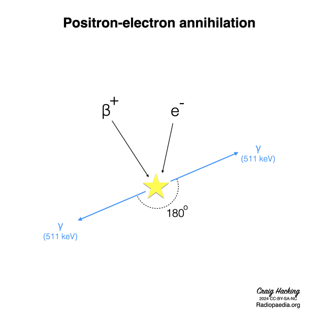
The positron-emitting isotope administered to the patient undergoes β+ decay in the body, with a proton being converted to a neutron, a positron (the antiparticle of the electron, sometimes referred to as a β+ particle), and a neutrino. The positron travels a short distance and annihilates with an electron. The annihilation reaction results in the formation of two high energy photons which travel in diametrically opposite directions.
Each photon has an energy of 511 keV. Two detectors at opposite ends facing each other detect these two photons traveling in opposite directions, and the radioactivity is localized somewhere along a line between the two detectors. This is referred to as the line of response.
Procedure
fasting for 4-6 hours - diabetic patients should fast overnight (12 hours)
blood glucose level <150 mg/dL
avoid strenuous activity 24 hours prior to imaging
avoid speech 20 minutes prior to imaging
the scan is carried out 60 minutes post-injection of FDG
In cases of fusion imaging such as PET-CT, the whole-body CT scan is conducted first, followed by the whole-body PET scan, and subsequently the two sets of images are co-registered.
A standardized uptake value (SUV) is calculated at the end of the study i.e. ratio of activity per unit mass tissue to injected dose per unit body mass. Other parameters include metabolic tumor volume (MTV) and total lesion glycolysis (TLG) 8.
Limitations
Motion artifacts result in an inaccurate anatomical co-registration of the CT and PET studies. The distance (2-3 mm) the positron travels before annihilation and the detector element size both contribute to relatively poor spatial resolution.
Physiological muscle uptake usually appears symmetrically and diffusely on PET imaging.
Attenuation correction
When gamma photons generated by radiopharmaceuticals pass through the body, it undergoes absorption and scatter, leading to attenuation of its signal 9. Therefore, when combined with CT, the CT imaging can be used to generate an attenuation map which is used to correct the PET imaging for attenuation. This attenuation correction can add a number of further artifacts due to errors in estimated the attenuation 9.
-
breathing
artifacts related to respiratory motion cause the 'mushroom effect' where an artifact is sometimes seen in the lung bases because of the different phases of respiratory motion
-
implants and prostheses
metallic implants such as joint prostheses can create significant artifact on PET images as the attenuation correction cannot deal with/correct for markedly high densities
-
truncation
CT field of view is limited whereas PET field of view is usually larger; if patients are scanned with arms by their side this can lead to the abnormal reconstruction of the images
Normal physiological uptake
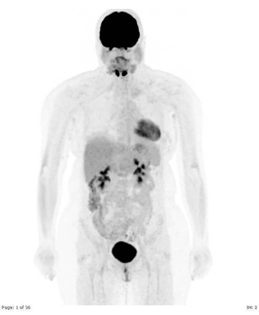
Waldeyer ring, e.g. palatine tonsils symmetrically, especially when younger 7
salivary glands
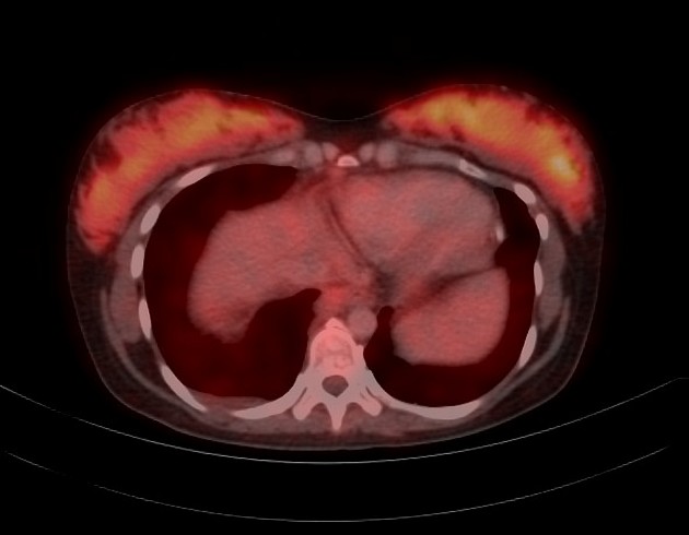
skeletal muscle, especially after strenuous activity and laryngeal muscles following speech
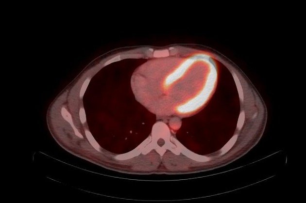
myocardium
gastrointestinal tract, e.g. intestinal wall
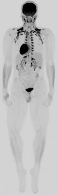
genitourinary tract: FDG is excreted via the renal system and passes into the collecting systems
thymus 4
lactating breasts
nipples
testicles
False-positive FDG uptake
This may occur due to the following conditions:
granulomatous disease
abscess
surgical changes
foreign body reaction e.g. talc pleurodesis
excessive bowel uptake with metformin therapy
inflammation (although at times e.g. evaluating for vasculitis, this may be the finding of interest)
Applications
-
oncologic
detection, staging, response to treatment
differentiation between radiation necrosis and recurrence
-
neurologic
early diagnosis of Alzheimer disease
localization of seizure focus in interictal phase
localizing eloquent areas (e.g. speech, motor function)
-
cardiac
identification of hibernating myocardium
-
infection/inflammation
History and etymology
David E Kuhl (1929-2017), a pioneering American nuclear physician, at the University of Pennsylvania, had a key role in the development of PET 6.


 Unable to process the form. Check for errors and try again.