
Accessory hemiazygos vein
Citation, DOI, disclosures and article data
At the time the article was created Henry Knipe had no recorded disclosures.
View Henry Knipe's current disclosuresAt the time the article was last revised Ian Bickle had the following disclosures:
- Hexarad, I work for this out sourcing company during non NHS hours (ongoing)
These were assessed during peer review and were determined to not be relevant to the changes that were made.
View Ian Bickle's current disclosures- Superior hemiazygos vein
- Accessory hemiazygos veins
- Superior hemiazygos veins
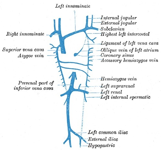
The accessory (or superior) hemiazygos vein forms part of the azygos system and along with the hemiazygos vein, it is partially analogous to the right-sided azygos vein. It drains the left superior hemithorax.
On this page:
Images:
Terminology
Spelling it "hemiazygous" when referring to the vein is incorrect, regardless of whether British or American English 5. In the context of anatomy, see Terminologia Anatomica, hemiazygos vein is the sole correct spelling.
Gross anatomy
Origin
The accessory hemiazygos vein is formed by the confluence of the 4th to 8th left posterior intercostal veins.
Course
It descends to the left of midline, adjacent to the thoracic vertebrae and crosses posteriorly to the aorta at the level of T7-8 to drain into the azygos vein. It normally anastomoses with the left superior intercostal vein superiorly.
Tributaries
left 4th-8th posterior intercostal veins
left bronchial veins (variable)
Variant anatomy
drains via a common trunk with the hemiazygos vein into the azygos vein
forms a common trunk with the hemiazygos vein that passes anterior to the aorta called the interazygos vein 2
drains directly into the left brachiocephalic vein (rare) 2
References
- 1. Ryan S, McNicholas M, Eustace SJ. Anatomy for Diagnostic Imaging. Saunders Ltd. ISBN:B006L65KZ2. Read it at Google Books - Find it at Amazon
- 2. Blackmon JM, Franco A. Normal variants of the accessory hemiazygos vein. Br J Radiol. 2011;84 (1003): 659-60. doi:10.1259/bjr/13695502 - Free text at pubmed - Pubmed citation
- 3. The vein book. Academic Press. ISBN:0123695155. Read it at Google Books - Find it at Amazon
- 4. Drake R, Vogl AW, Mitchell AWM. Gray's Basic Anatomy: with STUDENT CONSULT Online Access (Grays Anatomy for Students). Churchill Livingstone. ISBN:B0092DEQ1U. Read it at Google Books - Find it at Amazon
- 5. Holemans JA. Azygos, not azygous. (2001) AJR. American journal of roentgenology. 176 (6): 1602. doi:10.2214/ajr.176.6.1761602b - Pubmed
Incoming Links
- Bronchial vein
- Left upper lobe bronchus
- Thoracic duct
- Left main bronchus
- Absent azygos vein
- Left lower lobe
- Venous drainage of the thoracic wall
- Left lower lobe bronchus
- Left superior intercostal vein
- Subcostal artery
- Esophagus
- Left upper lobe
- Azygos vein
- Hemiazygos vein
- Azygos venous system
- Tracheobronchial tree
- Accessory hemiazygous vein
- Truncal venous development (Gray's illustration)
- Bronchiectasis and incidental accessory hemiazygos vein
- Aortic nipple
- Accessory hemiazygos vein draining into left brachiocephalic vein
- Superior vena cava invasion and obstruction
- Azygos venous system anatomy (CT pulmonary angiography)
- Left superior intercostal vein opacification
Related articles: Anatomy: Thoracic
- thoracic skeleton[+][+]
- thoracic cage
- thoracic spine
- articulations
- muscles of the thorax[+][+]
- diaphragm
- intercostal space
- intercostal muscles
- variant anatomy
- spaces of the thorax[+][+]
- thoracic viscera[+][+]
- lower respiratory tract
-
heart
- cardiac chambers
- heart valves
- cardiac fibrous skeleton
- innervation of the heart
- development of the heart
- cardiac wall
-
pericardium
- epicardium
- epicardial fat pad
- pericardial space
- oblique pericardial sinus
- transverse pericardial sinus
-
pericardial recesses
- aortic recesses
- pulmonic recesses
- postcaval recess
- pulmonary venous recesses
- pericardial ligaments
- myocardium
- endocardium
-
pericardium
- esophagus
- thymus
- breast
- arterial supply of the thorax[+][+]
-
thoracic aorta (development)
-
ascending aorta
-
aortic root
- aortic annulus
-
coronary arteries
- coronary arterial dominance
- myocardial segments
-
left main coronary artery (LMCA)
- ramus intermedius artery (RI)
-
circumflex artery (LCx)
- obtuse marginal branches (OM1, OM2, etc))
- Kugel's artery
-
left anterior descending artery (LAD)
- diagonal branches (D1, D2, etc)
- septal perforators (S1, S2, etc)
-
right coronary artery (RCA)
- conus artery
- sinoatrial nodal artery
- acute marginal branches (AM1, AM2, etc)
- inferior interventricular artery (PDA)
- posterior left ventricular artery (PLV)
- congenital anomalies
- sinotubular junction
-
aortic root
- aortic arch
- aortic isthmus
- descending aorta
-
ascending aorta
- pulmonary trunk
-
thoracic aorta (development)
- venous drainage of the thorax
- superior vena cava (SVC)
- inferior vena cava (IVC)[+][+]
-
coronary veins[+][+]
-
cardiac veins which drain into the coronary sinus
- great cardiac vein
- middle cardiac vein
- small cardiac vein
- posterior vein of the left ventricle
- vein of Marshall (oblique vein of the left atrium)
- anterior cardiac veins
- venae cordis minimae (smallest cardiac veins or thebesian veins)
-
cardiac veins which drain into the coronary sinus
- pulmonary veins
- bronchial veins
- thoracoepigastric vein
- lymphatics of the thorax[+][+]
- innervation of the thorax[+][+]


 Unable to process the form. Check for errors and try again.